

Advances in Neural Signal Processing
36
2.3 Experimental recording of gait using EEG
EEG was measured from four healthy subjects with four cycles per trial and two
trials were recorded per subject. All subjects had provided their informed consent
and were approved by the institutional ethical review board. Subjects were without
any known medical conditions and had normal or corrected-to-normal vision. All
subjects were explained the aim of the study before participating in the recordings.
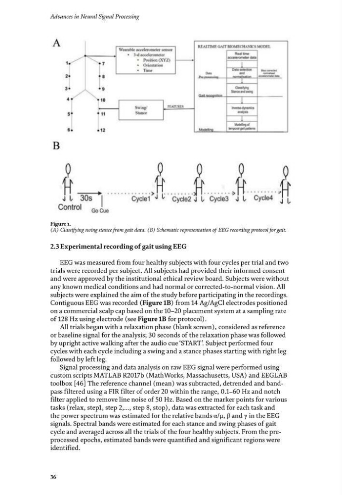
Contiguous EEG was recorded (Figure 1B) from 14 Ag/AgCl electrodes positioned
on a commercial scalp cap based on the 10–20 placement system at a sampling rate
of 128 Hz using electrode (see Figure 1B for protocol).
All trials began with a relaxation phase (blank screen), considered as reference
or baseline signal for the analysis; 30 seconds of the relaxation phase was followed
by upright active walking after the audio cue ‘START’ . Subject performed four
cycles with each cycle including a swing and a stance phases starting with right leg
followed by left leg.
Signal processing and data analysis on raw EEG signal were performed using
custom scripts MATLAB R2017b (MathWorks, Massachusetts, USA) and EEGLAB
toolbox [46] The reference channel (mean) was subtracted, detrended and bandpass filtered using a FIR filter of order 20 within the range, 0.1–60 Hz and notch
filter applied to remove line noise of 50 Hz. Based on the marker points for various
tasks (relax, step1, step 2,…, step 8, stop), data was extracted for each task and
the power spectrum was estimated for the relative bands α/μ, β and γ in the EEG
signals. Spectral bands were estimated for each stance and swing phases of gait
cycle and averaged across all the trials of the four healthy subjects. From the preprocessed epochs, estimated bands were quantified and significant regions were
identified.
Figure 1.
(A) Classifying swing stance from gait data. (B) Schematic representation of EEG recording protocol for gait.
36
2.3 Experimental recording of gait using EEG
EEG was measured from four healthy subjects with four cycles per trial and two
trials were recorded per subject. All subjects had provided their informed consent
and were approved by the institutional ethical review board. Subjects were without
any known medical conditions and had normal or corrected-to-normal vision. All
subjects were explained the aim of the study before participating in the recordings.
Contiguous EEG was recorded (Figure 1B) from 14 Ag/AgCl electrodes positioned
on a commercial scalp cap based on the 10–20 placement system at a sampling rate
of 128 Hz using electrode (see Figure 1B for protocol).
All trials began with a relaxation phase (blank screen), considered as reference
or baseline signal for the analysis; 30 seconds of the relaxation phase was followed
by upright active walking after the audio cue ‘START’ . Subject performed four
cycles with each cycle including a swing and a stance phases starting with right leg
followed by left leg.
Signal processing and data analysis on raw EEG signal were performed using
custom scripts MATLAB R2017b (MathWorks, Massachusetts, USA) and EEGLAB
toolbox [46] The reference channel (mean) was subtracted, detrended and bandpass filtered using a FIR filter of order 20 within the range, 0.1–60 Hz and notch
filter applied to remove line noise of 50 Hz. Based on the marker points for various
tasks (relax, step1, step 2,…, step 8, stop), data was extracted for each task and
the power spectrum was estimated for the relative bands α/μ, β and γ in the EEG
signals. Spectral bands were estimated for each stance and swing phases of gait
cycle and averaged across all the trials of the four healthy subjects. From the preprocessed epochs, estimated bands were quantified and significant regions were
identified.
Figure 1.
(A) Classifying swing stance from gait data. (B) Schematic representation of EEG recording protocol for gait.