

60
and functioning of mitochondria are reduced by reducing oxidative phosphorylation
[14]. In mammalian cells, as a key regulator of body functions, neurodegenerative
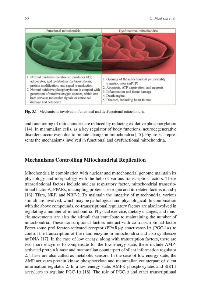
disorders occur even due to minute change in mitochondria [15]. Figure 3.1 represents the mechanisms involved in functional and dysfunctional mitochondria.
Mechanisms Controlling Mitochondrial Replication
Mitochondria in combination with nuclear and mitochondrial genome maintain its
physiology and morphology with the help of various transcription factors. These
transcriptional factors include nuclear respiratory factor, mitochondrial transcriptional factor A, PPARs, uncoupling proteins, estrogen and its related factors α and γ
[16], Tfam, NRF, and NRF-2. To maintain the integrity of mitochondria, various
stimuli are involved, which may be pathological and physiological. In combination
with the above compounds, co-transcriptional regulatory factors are also involved in
regulating a number of mitochondria. Physical exercise, dietary changes, and muscle movements are also the stimuli that contribute to maintaining the number of
mitochondria. These transcriptional factors interact with co-transcriptional factor
Peroxisome proliferator-activated receptor (PPAR)-γ coactivator-1α (PGC-1α) to
control the transcription of the main enzyme in mitochondria and also synthesize
mtDNA [17]. In the case of low energy, along with transcription factors, there are
two more enzymes to compensate for the low energy state, these include AMPactivated protein kinase and mammalian counterpart of silent information regulator
2. These are also called as metabolic sensors. In the case of low energy state, the
AMP activates protein kinase phosphorylate and mammalian counterpart of silent
information regulator 2. In a low energy state, AMPK phosphorylates and SIRT1
acetylates to regulate PGC-1α [18]. The role of PGC-α and other transcriptional
Fig. 3.1 Mechanisms involved in functional and dysfunctional mitochondria
G. Murtaza et al.
and functioning of mitochondria are reduced by reducing oxidative phosphorylation
[14]. In mammalian cells, as a key regulator of body functions, neurodegenerative
disorders occur even due to minute change in mitochondria [15]. Figure 3.1 represents the mechanisms involved in functional and dysfunctional mitochondria.
Mechanisms Controlling Mitochondrial Replication
Mitochondria in combination with nuclear and mitochondrial genome maintain its
physiology and morphology with the help of various transcription factors. These
transcriptional factors include nuclear respiratory factor, mitochondrial transcriptional factor A, PPARs, uncoupling proteins, estrogen and its related factors α and γ
[16], Tfam, NRF, and NRF-2. To maintain the integrity of mitochondria, various
stimuli are involved, which may be pathological and physiological. In combination
with the above compounds, co-transcriptional regulatory factors are also involved in
regulating a number of mitochondria. Physical exercise, dietary changes, and muscle movements are also the stimuli that contribute to maintaining the number of
mitochondria. These transcriptional factors interact with co-transcriptional factor
Peroxisome proliferator-activated receptor (PPAR)-γ coactivator-1α (PGC-1α) to
control the transcription of the main enzyme in mitochondria and also synthesize
mtDNA [17]. In the case of low energy, along with transcription factors, there are
two more enzymes to compensate for the low energy state, these include AMPactivated protein kinase and mammalian counterpart of silent information regulator
2. These are also called as metabolic sensors. In the case of low energy state, the
AMP activates protein kinase phosphorylate and mammalian counterpart of silent
information regulator 2. In a low energy state, AMPK phosphorylates and SIRT1
acetylates to regulate PGC-1α [18]. The role of PGC-α and other transcriptional
Fig. 3.1 Mechanisms involved in functional and dysfunctional mitochondria
G. Murtaza et al.