

general agreement with two previous studies that showed reduced expression of
maturation markers and proinflammatory cytokines following DC exposure to
piperine (Bae et al. 2012); however, unlike these earlier reports, we observed
reduced synthesis of IL-6, but no effect on CD86 expression by piperine-treated
DCs. Reduced production of proinflammatory cytokines has also been reported in
piperine-treated B16-F10 melanoma cells, adipocytes, and macrophages (Pradeep
and Kuttan 2003, 2004; Woo et al. 2007) indicating that this effect is not cell
type-specific. Moreover, normal expression of ICAM-1, MAC-1, and LFA-1
adhesion molecules by piperine-treated DCs shows selective inhibition of DC
surface marker expression, which may reflect targeting of specific signal transduction pathways. Indeed, piperine is a known inhibitor of extracellular
signal-regulated and c-Jun N-terminal kinases that are activated in mouse DCs as a
result of LPS stimulation (Bae et al. 2012).
Piperine-mediated inhibition of DC maturation marker expression induced by
LPS, CpG, and poly I:C indicates that the inhibitory effect of piperine on DC maturation caused by pattern recognition receptor stimulation was not restricted to the
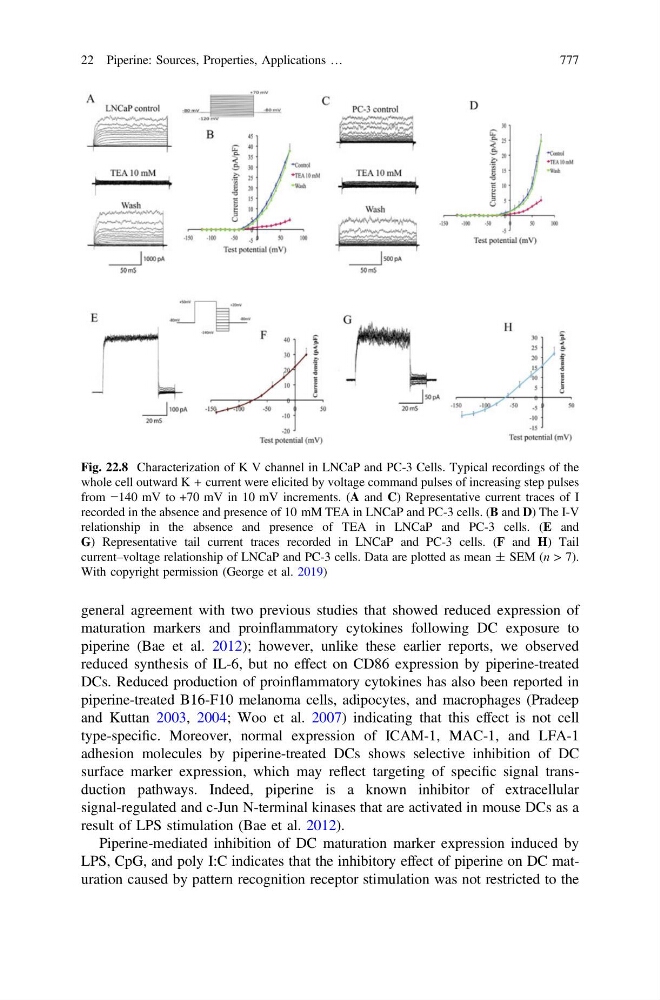
Fig. 22.8 Characterization of K V channel in LNCaP and PC-3 Cells. Typical recordings of the
whole cell outward K + current were elicited by voltage command pulses of increasing step pulses
from −140 mV to +70 mV in 10 mV increments. (A and C) Representative current traces of I
recorded in the absence and presence of 10 mM TEA in LNCaP and PC-3 cells. (B and D) The I-V
relationship in the absence and presence of TEA in LNCaP and PC-3 cells. (E and
G) Representative tail current traces recorded in LNCaP and PC-3 cells. (F and H) Tail
current–voltage relationship of LNCaP and PC-3 cells. Data are plotted as mean ± SEM (n > 7).
With copyright permission (George et al. 2019)
22 Piperine: Sources, Properties, Applications …
777