

while in a concentration range of 0.1 ng/mL–10 µg/mL did not exhibit any kind of
inhibition in the cell growth of human foreskin fibroblast cells (HF cells, normal
cells).
Lim et al. isolated 17 alkaloids (130–146) from the ethanolic extract of A.
macrophylla stem bark and leaf (Fig. 14.31). Further, they evaluated the MDR
reversing
capacity
in
KB/S
(vincristine-sensitive)
and
KB/VJ300
(vincristine-sensitive and vincristine-resistant KB cell lines) (Lim et al. 2014).
All the compounds exhibited poor cytotoxicity in the cell lines (IC 50 > 25 µg/mL).
However, 11-methoxyvincorine (141), 11-demethoxyquaternine (144), 19,20-Zaffinisine (136) were reported to reverse MDR in vincristine-resistant KB (VJ300)
cells (IC 50 = 4.60, 6.60 and 7.47 µg/mL, respectively).
14.3.2.2 Antiplasmodial Activity
Keawpradub et al. isolated 13 alkaloids (114–116, 118–128) from the methanolic
extract of A. macrophylla leaves, stem bark, root bark and evaluated the antiplasmodial activity against the K1 strain of P. falciparum by using chloroquine
diphosphate as a positive control (Keawpradub et al. 1999). Bisindole alkaloids
such as 114, 115, 116, and 128 (IC 50 values = 0.53, 0.36, 0.27, and 1.12 µM)
exhibited a comparable activity with the chloroquine diphosphate (IC 50
value = 0.20 µM). Further, the effects of these alkaloids were evaluated in
chloroquine-resistant (K1) and chloroquine-sensitive strains (T9–96) of
P. falciparum. These bisindoles were found to be significantly less active against
T9–96 (0.94–39 µM; chloroquine diphosphate—0.02 µM). In K1 cells, they
exhibited a significant activity (0.36–1.12 µM; chloroquine diphosphate—
0.20 µM).
Hirasawa et al. isolated alstiphyllanines A-D (147–150, Fig. 14.32) from the
methanolic extract of A. macrophylla leaves and evaluated the antiplasmodial
activity (against P. falciparum, 3D7) (Hirasawa et al. 2009). Screened alkaloids
exhibited a moderate in vitro antiplasmodial activity (IC 50 of 6.85, 0.34, 6.20, and
2.75 µg/mL, respectively, for 147–150). In vitro cytotoxicity evaluation resulted in
O
O
O
O
O OH
HO
HO
O
O
O
O
O
HO
HO
OH
O
O
H
H
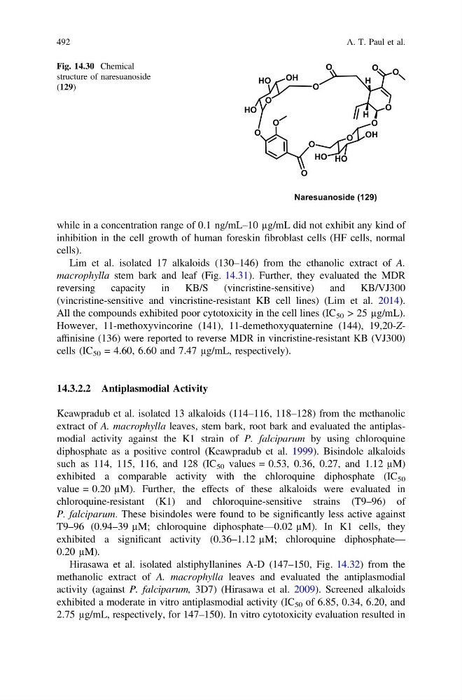
Naresuanoside (129)
Fig. 14.30 Chemical
structure of naresuanoside
(129)
492
A. T. Paul et al.
inhibition in the cell growth of human foreskin fibroblast cells (HF cells, normal
cells).
Lim et al. isolated 17 alkaloids (130–146) from the ethanolic extract of A.
macrophylla stem bark and leaf (Fig. 14.31). Further, they evaluated the MDR
reversing
capacity
in
KB/S
(vincristine-sensitive)
and
KB/VJ300
(vincristine-sensitive and vincristine-resistant KB cell lines) (Lim et al. 2014).
All the compounds exhibited poor cytotoxicity in the cell lines (IC 50 > 25 µg/mL).
However, 11-methoxyvincorine (141), 11-demethoxyquaternine (144), 19,20-Zaffinisine (136) were reported to reverse MDR in vincristine-resistant KB (VJ300)
cells (IC 50 = 4.60, 6.60 and 7.47 µg/mL, respectively).
14.3.2.2 Antiplasmodial Activity
Keawpradub et al. isolated 13 alkaloids (114–116, 118–128) from the methanolic
extract of A. macrophylla leaves, stem bark, root bark and evaluated the antiplasmodial activity against the K1 strain of P. falciparum by using chloroquine
diphosphate as a positive control (Keawpradub et al. 1999). Bisindole alkaloids
such as 114, 115, 116, and 128 (IC 50 values = 0.53, 0.36, 0.27, and 1.12 µM)
exhibited a comparable activity with the chloroquine diphosphate (IC 50
value = 0.20 µM). Further, the effects of these alkaloids were evaluated in
chloroquine-resistant (K1) and chloroquine-sensitive strains (T9–96) of
P. falciparum. These bisindoles were found to be significantly less active against
T9–96 (0.94–39 µM; chloroquine diphosphate—0.02 µM). In K1 cells, they
exhibited a significant activity (0.36–1.12 µM; chloroquine diphosphate—
0.20 µM).
Hirasawa et al. isolated alstiphyllanines A-D (147–150, Fig. 14.32) from the
methanolic extract of A. macrophylla leaves and evaluated the antiplasmodial
activity (against P. falciparum, 3D7) (Hirasawa et al. 2009). Screened alkaloids
exhibited a moderate in vitro antiplasmodial activity (IC 50 of 6.85, 0.34, 6.20, and
2.75 µg/mL, respectively, for 147–150). In vitro cytotoxicity evaluation resulted in
O
O
O
O
O OH
HO
HO
O
O
O
O
O
HO
HO
OH
O
O
H
H
Naresuanoside (129)
Fig. 14.30 Chemical
structure of naresuanoside
(129)
492
A. T. Paul et al.