

180
R. Vanparys et al.
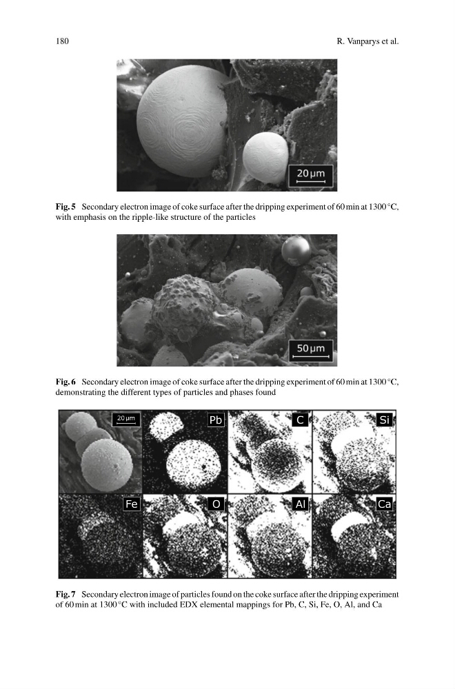
Fig. 5 Secondary electron image of coke surface after the dripping experiment of 60 min at 1300 °C,
with emphasis on the ripple-like structure of the particles
Fig. 6 Secondary electron image of coke surface after the dripping experiment of 60 min at 1300 °C,
demonstrating the different types of particles and phases found
µ
Fig. 7 Secondary electron image of particles found on the coke surface after the dripping experiment
of 60 min at 1300 °C with included EDX elemental mappings for Pb, C, Si, Fe, O, Al, and Ca
R. Vanparys et al.
Fig. 5 Secondary electron image of coke surface after the dripping experiment of 60 min at 1300 °C,
with emphasis on the ripple-like structure of the particles
Fig. 6 Secondary electron image of coke surface after the dripping experiment of 60 min at 1300 °C,
demonstrating the different types of particles and phases found
µ
Fig. 7 Secondary electron image of particles found on the coke surface after the dripping experiment
of 60 min at 1300 °C with included EDX elemental mappings for Pb, C, Si, Fe, O, Al, and Ca