

36
D. P. Barai et al.
different terminologies even though the latter is dependent on the earlier. There are
many factors that affect the particle size of the nanoparticles in the nanofluid and so
they indirectly affect the particle size distribution.
5.1 Characterization Techniques Used for Particle Size
Distribution
There are several methods that can be used for determining the particles size of the
nanoparticles dispersed in the nanofluid and characterize its particle size distribution
(Lin et al. 2014). Dynamic light scattering is the method in which the intensities at
which the laser beam is scattered by the nanoparticles are measured, which depends
on their Brownian motion in the nanofluid. So, the hydrodynamic diameter (D h ) as
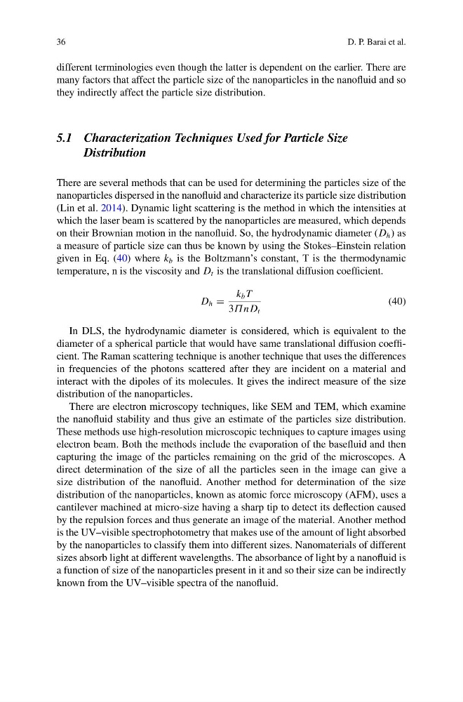
a measure of particle size can thus be known by using the Stokes–Einstein relation
given in Eq. (40) where k b is the Boltzmann’s constant, T is the thermodynamic
temperature, n is the viscosity and D t is the translational diffusion coefficient.
D h =
k b T
3Π n D t
(40)
In DLS, the hydrodynamic diameter is considered, which is equivalent to the
diameter of a spherical particle that would have same translational diffusion coefficient. The Raman scattering technique is another technique that uses the differences
in frequencies of the photons scattered after they are incident on a material and
interact with the dipoles of its molecules. It gives the indirect measure of the size
distribution of the nanoparticles.
There are electron microscopy techniques, like SEM and TEM, which examine
the nanofluid stability and thus give an estimate of the particles size distribution.
These methods use high-resolution microscopic techniques to capture images using
electron beam. Both the methods include the evaporation of the basefluid and then
capturing the image of the particles remaining on the grid of the microscopes. A
direct determination of the size of all the particles seen in the image can give a
size distribution of the nanofluid. Another method for determination of the size
distribution of the nanoparticles, known as atomic force microscopy (AFM), uses a
cantilever machined at micro-size having a sharp tip to detect its deflection caused
by the repulsion forces and thus generate an image of the material. Another method
is the UV–visible spectrophotometry that makes use of the amount of light absorbed
by the nanoparticles to classify them into different sizes. Nanomaterials of different
sizes absorb light at different wavelengths. The absorbance of light by a nanofluid is
a function of size of the nanoparticles present in it and so their size can be indirectly
known from the UV–visible spectra of the nanofluid.
D. P. Barai et al.
different terminologies even though the latter is dependent on the earlier. There are
many factors that affect the particle size of the nanoparticles in the nanofluid and so
they indirectly affect the particle size distribution.
5.1 Characterization Techniques Used for Particle Size
Distribution
There are several methods that can be used for determining the particles size of the
nanoparticles dispersed in the nanofluid and characterize its particle size distribution
(Lin et al. 2014). Dynamic light scattering is the method in which the intensities at
which the laser beam is scattered by the nanoparticles are measured, which depends
on their Brownian motion in the nanofluid. So, the hydrodynamic diameter (D h ) as
a measure of particle size can thus be known by using the Stokes–Einstein relation
given in Eq. (40) where k b is the Boltzmann’s constant, T is the thermodynamic
temperature, n is the viscosity and D t is the translational diffusion coefficient.
D h =
k b T
3Π n D t
(40)
In DLS, the hydrodynamic diameter is considered, which is equivalent to the
diameter of a spherical particle that would have same translational diffusion coefficient. The Raman scattering technique is another technique that uses the differences
in frequencies of the photons scattered after they are incident on a material and
interact with the dipoles of its molecules. It gives the indirect measure of the size
distribution of the nanoparticles.
There are electron microscopy techniques, like SEM and TEM, which examine
the nanofluid stability and thus give an estimate of the particles size distribution.
These methods use high-resolution microscopic techniques to capture images using
electron beam. Both the methods include the evaporation of the basefluid and then
capturing the image of the particles remaining on the grid of the microscopes. A
direct determination of the size of all the particles seen in the image can give a
size distribution of the nanofluid. Another method for determination of the size
distribution of the nanoparticles, known as atomic force microscopy (AFM), uses a
cantilever machined at micro-size having a sharp tip to detect its deflection caused
by the repulsion forces and thus generate an image of the material. Another method
is the UV–visible spectrophotometry that makes use of the amount of light absorbed
by the nanoparticles to classify them into different sizes. Nanomaterials of different
sizes absorb light at different wavelengths. The absorbance of light by a nanofluid is
a function of size of the nanoparticles present in it and so their size can be indirectly
known from the UV–visible spectra of the nanofluid.