

Microstructurally Engineered Ceramics for Environmental …
517
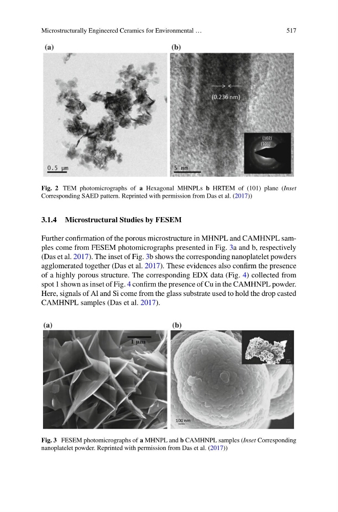
Fig. 2 TEM photomicrographs of a Hexagonal MHNPLs b HRTEM of (101) plane (Inset
Corresponding SAED pattern. Reprinted with permission from Das et al. (2017))
3.1.4 Microstructural Studies by FESEM
Further confirmation of the porous microstructure in MHNPL and CAMHNPL samples come from FESEM photomicrographs presented in Fig. 3a and b, respectively
(Das et al. 2017). The inset of Fig. 3b shows the corresponding nanoplatelet powders
agglomerated together (Das et al. 2017). These evidences also confirm the presence
of a highly porous structure. The corresponding EDX data (Fig. 4) collected from
spot 1 shown as inset of Fig. 4 confirm the presence of Cu in the CAMHNPL powder.
Here, signals of Al and Si come from the glass substrate used to hold the drop casted
CAMHNPL samples (Das et al. 2017).
Fig. 3 FESEM photomicrographs of a MHNPL and b CAMHNPL samples (Inset Corresponding
nanoplatelet powder. Reprinted with permission from Das et al. (2017))
517
Fig. 2 TEM photomicrographs of a Hexagonal MHNPLs b HRTEM of (101) plane (Inset
Corresponding SAED pattern. Reprinted with permission from Das et al. (2017))
3.1.4 Microstructural Studies by FESEM
Further confirmation of the porous microstructure in MHNPL and CAMHNPL samples come from FESEM photomicrographs presented in Fig. 3a and b, respectively
(Das et al. 2017). The inset of Fig. 3b shows the corresponding nanoplatelet powders
agglomerated together (Das et al. 2017). These evidences also confirm the presence
of a highly porous structure. The corresponding EDX data (Fig. 4) collected from
spot 1 shown as inset of Fig. 4 confirm the presence of Cu in the CAMHNPL powder.
Here, signals of Al and Si come from the glass substrate used to hold the drop casted
CAMHNPL samples (Das et al. 2017).
Fig. 3 FESEM photomicrographs of a MHNPL and b CAMHNPL samples (Inset Corresponding
nanoplatelet powder. Reprinted with permission from Das et al. (2017))