

514
P. S. Das et al.
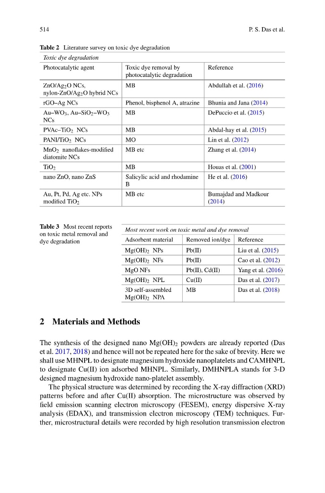
Table 2 Literature survey on toxic dye degradation
Toxic dye degradation
Photocatalytic agent
Toxic dye removal by
photocatalytic degradation
Reference
ZnO/Ag 2 O NCs,
nylon-ZnO/Ag 2 O hybrid NCs
MB
Abdullah et al. (2016)
rGO–Ag NCs
Phenol, bisphenol A, atrazine Bhunia and Jana (2014)
Au–WO 3 , Au–SiO 2 –WO 3
NCs
MB
DePuccio et al. (2015)
PVAc–TiO 2 NCs
MB
Abdal-hay et al. (2015)
PANI/TiO 2 NCs
MO
Lin et al. (2012)
MnO 2 nanoflakes-modified
diatomite NCs
MB etc
Zhang et al. (2014)
TiO 2
MB
Houas et al. (2001)
nano ZnO, nano ZnS
Salicylic acid and rhodamine
B
He et al. (2016)
Au, Pt, Pd, Ag etc. NPs
modified TiO 2
MB etc
Bumajdad and Madkour
(2014)
Table 3 Most recent reports
on toxic metal removal and
dye degradation
Most recent work on toxic metal and dye removal
Adsorbent material
Removed ion/dye Reference
Mg(OH) 2 NPs
Pb(II)
Liu et al. (2015)
Mg(OH) 2 NFs
Pb(II)
Cao et al. (2012)
MgO NFs
Pb(II), Cd(II)
Yang et al. (2016)
Mg(OH) 2 NPL
Cu(II)
Das et al. (2017)
3D self-assembled
Mg(OH) 2 NPA
MB
Das et al. (2018)
2 Materials and Methods
The synthesis of the designed nano Mg(OH) 2 powders are already reported (Das
et al. 2017, 2018) and hence will not be repeated here for the sake of brevity. Here we
shall use MHNPL to designate magnesium hydroxide nanoplatelets and CAMHNPL
to designate Cu(II) ion adsorbed MHNPL. Similarly, DMHNPLA stands for 3-D
designed magnesium hydroxide nano-platelet assembly.
The physical structure was determined by recording the X-ray diffraction (XRD)
patterns before and after Cu(II) absorption. The microstructure was observed by
field emission scanning electron microscopy (FESEM), energy dispersive X-ray
analysis (EDAX), and transmission electron microscopy (TEM) techniques. Further, microstructural details were recorded by high resolution transmission electron
P. S. Das et al.
Table 2 Literature survey on toxic dye degradation
Toxic dye degradation
Photocatalytic agent
Toxic dye removal by
photocatalytic degradation
Reference
ZnO/Ag 2 O NCs,
nylon-ZnO/Ag 2 O hybrid NCs
MB
Abdullah et al. (2016)
rGO–Ag NCs
Phenol, bisphenol A, atrazine Bhunia and Jana (2014)
Au–WO 3 , Au–SiO 2 –WO 3
NCs
MB
DePuccio et al. (2015)
PVAc–TiO 2 NCs
MB
Abdal-hay et al. (2015)
PANI/TiO 2 NCs
MO
Lin et al. (2012)
MnO 2 nanoflakes-modified
diatomite NCs
MB etc
Zhang et al. (2014)
TiO 2
MB
Houas et al. (2001)
nano ZnO, nano ZnS
Salicylic acid and rhodamine
B
He et al. (2016)
Au, Pt, Pd, Ag etc. NPs
modified TiO 2
MB etc
Bumajdad and Madkour
(2014)
Table 3 Most recent reports
on toxic metal removal and
dye degradation
Most recent work on toxic metal and dye removal
Adsorbent material
Removed ion/dye Reference
Mg(OH) 2 NPs
Pb(II)
Liu et al. (2015)
Mg(OH) 2 NFs
Pb(II)
Cao et al. (2012)
MgO NFs
Pb(II), Cd(II)
Yang et al. (2016)
Mg(OH) 2 NPL
Cu(II)
Das et al. (2017)
3D self-assembled
Mg(OH) 2 NPA
MB
Das et al. (2018)
2 Materials and Methods
The synthesis of the designed nano Mg(OH) 2 powders are already reported (Das
et al. 2017, 2018) and hence will not be repeated here for the sake of brevity. Here we
shall use MHNPL to designate magnesium hydroxide nanoplatelets and CAMHNPL
to designate Cu(II) ion adsorbed MHNPL. Similarly, DMHNPLA stands for 3-D
designed magnesium hydroxide nano-platelet assembly.
The physical structure was determined by recording the X-ray diffraction (XRD)
patterns before and after Cu(II) absorption. The microstructure was observed by
field emission scanning electron microscopy (FESEM), energy dispersive X-ray
analysis (EDAX), and transmission electron microscopy (TEM) techniques. Further, microstructural details were recorded by high resolution transmission electron