

116
A. Bagaria and S. Ramakumar
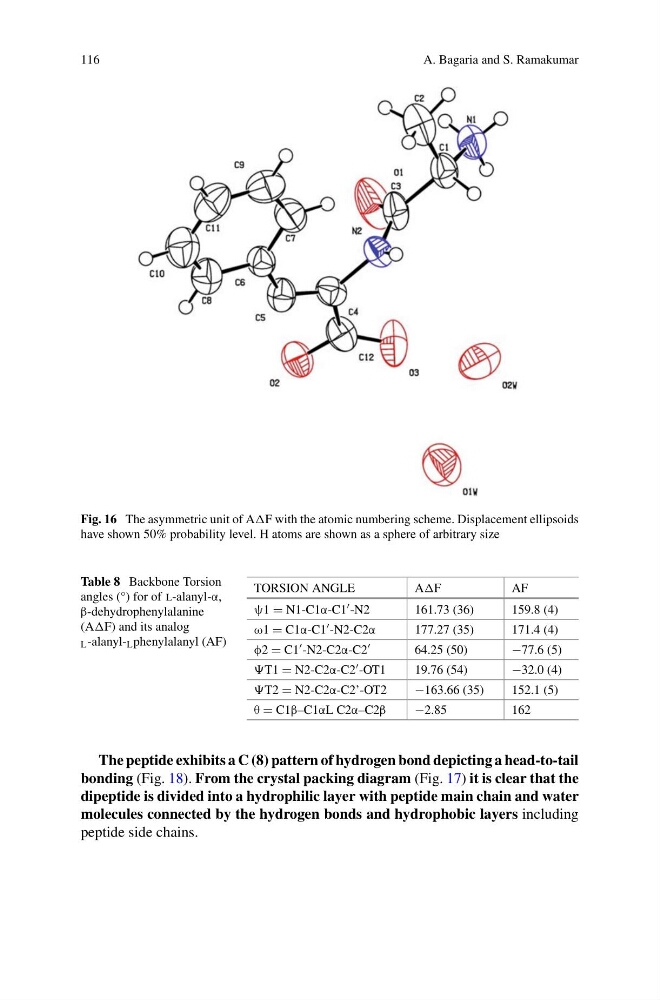
Fig. 16 The asymmetric unit of AF with the atomic numbering scheme. Displacement ellipsoids
have shown 50% probability level. H atoms are shown as a sphere of arbitrary size
Table 8 Backbone Torsion
angles (°) for of l-alanyl-α,
β-dehydrophenylalanine
(AF) and its analog
L -alanyl- L phenylalanyl (AF)
TORSION ANGLE
AF
AF
ψ1 = N1-C1α-C1 -N2
161.73 (36)
159.8 (4)
ω1 = C1α-C1 -N2-C2α
177.27 (35)
171.4 (4)
φ2 = C1 -N2-C2α-C2
64.25 (50)
−77.6 (5)
T1 = N2-C2α-C2 -OT1
19.76 (54)
−32.0 (4)
T2 = N2-C2α-C2’-OT2
−163.66 (35)
152.1 (5)
θ = C1β–C1αL C2α–C2β
−2.85
162
The peptide exhibits a C (8) pattern of hydrogen bond depicting a head-to-tail
bonding (Fig. 18). From the crystal packing diagram (Fig. 17) it is clear that the
dipeptide is divided into a hydrophilic layer with peptide main chain and water
molecules connected by the hydrogen bonds and hydrophobic layers including
peptide side chains.
A. Bagaria and S. Ramakumar
Fig. 16 The asymmetric unit of AF with the atomic numbering scheme. Displacement ellipsoids
have shown 50% probability level. H atoms are shown as a sphere of arbitrary size
Table 8 Backbone Torsion
angles (°) for of l-alanyl-α,
β-dehydrophenylalanine
(AF) and its analog
L -alanyl- L phenylalanyl (AF)
TORSION ANGLE
AF
AF
ψ1 = N1-C1α-C1 -N2
161.73 (36)
159.8 (4)
ω1 = C1α-C1 -N2-C2α
177.27 (35)
171.4 (4)
φ2 = C1 -N2-C2α-C2
64.25 (50)
−77.6 (5)
T1 = N2-C2α-C2 -OT1
19.76 (54)
−32.0 (4)
T2 = N2-C2α-C2’-OT2
−163.66 (35)
152.1 (5)
θ = C1β–C1αL C2α–C2β
−2.85
162
The peptide exhibits a C (8) pattern of hydrogen bond depicting a head-to-tail
bonding (Fig. 18). From the crystal packing diagram (Fig. 17) it is clear that the
dipeptide is divided into a hydrophilic layer with peptide main chain and water
molecules connected by the hydrogen bonds and hydrophobic layers including
peptide side chains.