

Peptide Nanotubes: A Crystallographic Approach
105
Various hydrogen bonds and the parameters for the Phe-Phe molecule are tabulated in Table 3. The crystal structure of the Phe–Phe dipeptide shows a C(8)
pattern (Etter et al. 1990) of head-to-tail hydrogen bonding meaning that eight atoms
are encountered in traversing the shortest pathway from the hydrogen atom of one
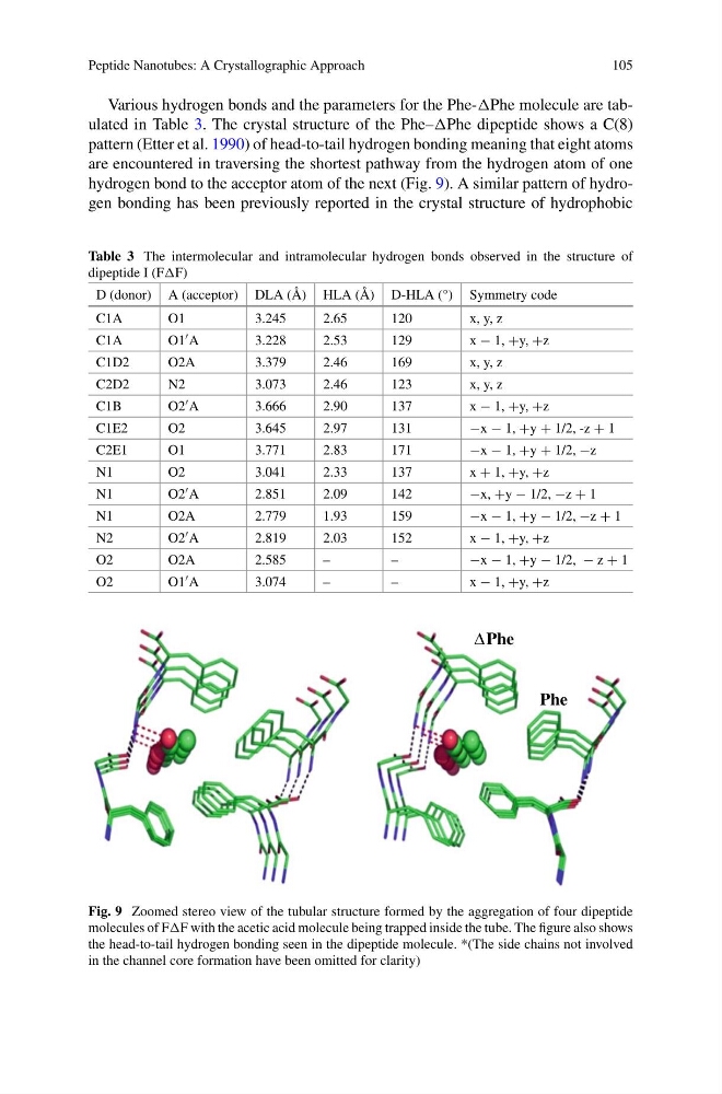
hydrogen bond to the acceptor atom of the next (Fig. 9). A similar pattern of hydrogen bonding has been previously reported in the crystal structure of hydrophobic
Table 3 The intermolecular and intramolecular hydrogen bonds observed in the structure of
dipeptide I (FF)
D (donor) A (acceptor) DLA (Å) HLA (Å) D-HLA (°) Symmetry code
C1A
O1
3.245
2.65
120
x, y, z
C1A
O1 A
3.228
2.53
129
x − 1, +y, +z
C1D2
O2A
3.379
2.46
169
x, y, z
C2D2
N2
3.073
2.46
123
x, y, z
C1B
O2 A
3.666
2.90
137
x − 1, +y, +z
C1E2
O2
3.645
2.97
131
−x − 1, +y + 1/2, -z + 1
C2E1
O1
3.771
2.83
171
−x − 1, +y + 1/2, −z
N1
O2
3.041
2.33
137
x + 1, +y, +z
N1
O2 A
2.851
2.09
142
−x, +y − 1/2, −z + 1
N1
O2A
2.779
1.93
159
−x − 1, +y − 1/2, −z + 1
N2
O2 A
2.819
2.03
152
x − 1, +y, +z
O2
O2A
2.585
–
–
−x − 1, +y − 1/2, − z + 1
O2
O1 A
3.074
–
–
x − 1, +y, +z
Phe
ΔPhe
Fig. 9 Zoomed stereo view of the tubular structure formed by the aggregation of four dipeptide
molecules of FF with the acetic acid molecule being trapped inside the tube. The figure also shows
the head-to-tail hydrogen bonding seen in the dipeptide molecule. *(The side chains not involved
in the channel core formation have been omitted for clarity)
105
Various hydrogen bonds and the parameters for the Phe-Phe molecule are tabulated in Table 3. The crystal structure of the Phe–Phe dipeptide shows a C(8)
pattern (Etter et al. 1990) of head-to-tail hydrogen bonding meaning that eight atoms
are encountered in traversing the shortest pathway from the hydrogen atom of one
hydrogen bond to the acceptor atom of the next (Fig. 9). A similar pattern of hydrogen bonding has been previously reported in the crystal structure of hydrophobic
Table 3 The intermolecular and intramolecular hydrogen bonds observed in the structure of
dipeptide I (FF)
D (donor) A (acceptor) DLA (Å) HLA (Å) D-HLA (°) Symmetry code
C1A
O1
3.245
2.65
120
x, y, z
C1A
O1 A
3.228
2.53
129
x − 1, +y, +z
C1D2
O2A
3.379
2.46
169
x, y, z
C2D2
N2
3.073
2.46
123
x, y, z
C1B
O2 A
3.666
2.90
137
x − 1, +y, +z
C1E2
O2
3.645
2.97
131
−x − 1, +y + 1/2, -z + 1
C2E1
O1
3.771
2.83
171
−x − 1, +y + 1/2, −z
N1
O2
3.041
2.33
137
x + 1, +y, +z
N1
O2 A
2.851
2.09
142
−x, +y − 1/2, −z + 1
N1
O2A
2.779
1.93
159
−x − 1, +y − 1/2, −z + 1
N2
O2 A
2.819
2.03
152
x − 1, +y, +z
O2
O2A
2.585
–
–
−x − 1, +y − 1/2, − z + 1
O2
O1 A
3.074
–
–
x − 1, +y, +z
Phe
ΔPhe
Fig. 9 Zoomed stereo view of the tubular structure formed by the aggregation of four dipeptide
molecules of FF with the acetic acid molecule being trapped inside the tube. The figure also shows
the head-to-tail hydrogen bonding seen in the dipeptide molecule. *(The side chains not involved
in the channel core formation have been omitted for clarity)