

104
A. Bagaria and S. Ramakumar
Table 2 Backbone Torsion
angles (°) for
L -Phenyalanyl-α,
β-dehydrophenylalanine,
dipeptide I (FF) and L -
Phenyalanyl- L -phenylalanine
(FF)
Torsion angle
FF
FF
ψ 1 = N 1 –C α
1 –C 1
–N 2
150.57 (20)
157.82 (4)
ω1 = C α
1 –C 1
–N 2
C α
2
177.96 (20)
−179.10 (4)
φ2 = C 1
–N 2 –C α
2 –C 2
−66.84 (28)
55.40 (5)
T1 = N 2 –C α
2 –C 2
–OT1
−29.3 (3)
43.8 (5)
T2 = N 2 –C α
2 –C 2
-OT2
154.1 (2)
−139.8 (4)
θ = C
β
1 –C α
1 L C α
2 –C
β
2
149.70
40.21
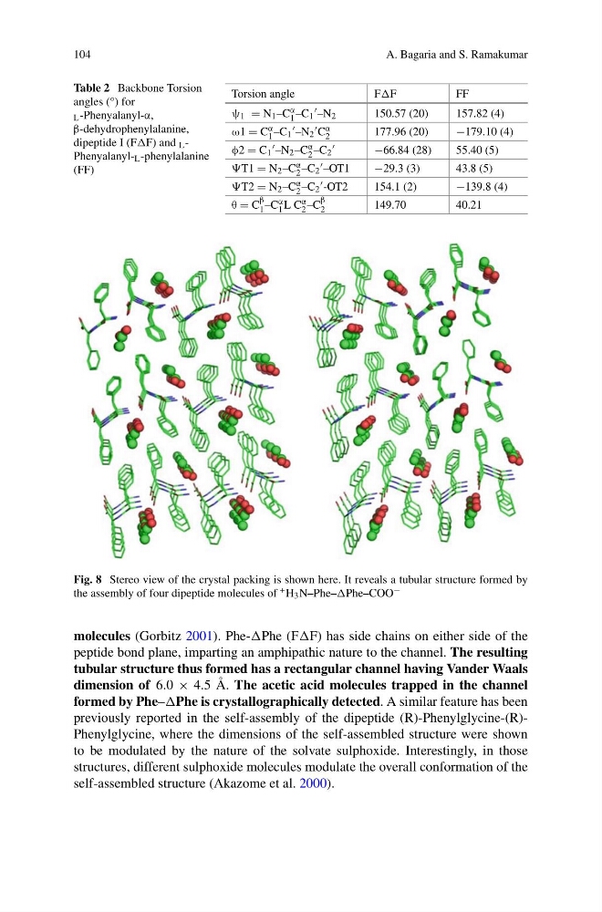
Fig. 8 Stereo view of the crystal packing is shown here. It reveals a tubular structure formed by
the assembly of four dipeptide molecules of + H 3 N–Phe–Phe–COO −
molecules (Gorbitz 2001). Phe-Phe (FF) has side chains on either side of the
peptide bond plane, imparting an amphipathic nature to the channel. The resulting
tubular structure thus formed has a rectangular channel having Vander Waals
dimension of 6.0 × 4.5 Å. The acetic acid molecules trapped in the channel
formed by Phe–Phe is crystallographically detected. A similar feature has been
previously reported in the self-assembly of the dipeptide (R)-Phenylglycine-(R)Phenylglycine, where the dimensions of the self-assembled structure were shown
to be modulated by the nature of the solvate sulphoxide. Interestingly, in those
structures, different sulphoxide molecules modulate the overall conformation of the
self-assembled structure (Akazome et al. 2000).
A. Bagaria and S. Ramakumar
Table 2 Backbone Torsion
angles (°) for
L -Phenyalanyl-α,
β-dehydrophenylalanine,
dipeptide I (FF) and L -
Phenyalanyl- L -phenylalanine
(FF)
Torsion angle
FF
FF
ψ 1 = N 1 –C α
1 –C 1
–N 2
150.57 (20)
157.82 (4)
ω1 = C α
1 –C 1
–N 2
C α
2
177.96 (20)
−179.10 (4)
φ2 = C 1
–N 2 –C α
2 –C 2
−66.84 (28)
55.40 (5)
T1 = N 2 –C α
2 –C 2
–OT1
−29.3 (3)
43.8 (5)
T2 = N 2 –C α
2 –C 2
-OT2
154.1 (2)
−139.8 (4)
θ = C
β
1 –C α
1 L C α
2 –C
β
2
149.70
40.21
Fig. 8 Stereo view of the crystal packing is shown here. It reveals a tubular structure formed by
the assembly of four dipeptide molecules of + H 3 N–Phe–Phe–COO −
molecules (Gorbitz 2001). Phe-Phe (FF) has side chains on either side of the
peptide bond plane, imparting an amphipathic nature to the channel. The resulting
tubular structure thus formed has a rectangular channel having Vander Waals
dimension of 6.0 × 4.5 Å. The acetic acid molecules trapped in the channel
formed by Phe–Phe is crystallographically detected. A similar feature has been
previously reported in the self-assembly of the dipeptide (R)-Phenylglycine-(R)Phenylglycine, where the dimensions of the self-assembled structure were shown
to be modulated by the nature of the solvate sulphoxide. Interestingly, in those
structures, different sulphoxide molecules modulate the overall conformation of the
self-assembled structure (Akazome et al. 2000).