

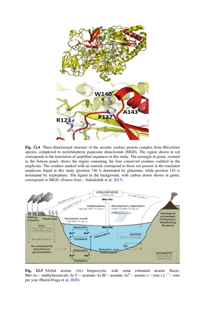
Fig. 12.4 Three-dimensional structure of the arsenite oxidase protein complex from Rhizobium
species, complexed to molybdopterin guanosine dinucleotide (MGD). The region shown in red
corresponds to the translation of amplified sequences in this study. The rectangle in green, zoomed
in the bottom panel, shows the region containing the four conserved residues codified in the
amplicons. The residues marked with an asterisk correspond to those not present in the translated
amplicons found in this study (position 140 is dominated by glutamate, while position 143 is
dominated by tryptophan). The ligand in the background, with carbon atoms shown in green,
corresponds to MGD. (Source from – Suhadolnik et al. 2017)
Fig. 12.5 Global arsenic (As) biogeocycle, with some estimated arsenic fluxes.
Met-As ¼ methylarsenicals; As V ¼ arsenate; As III ¼ arsenite; As
0 ¼ arsenic; t ¼ tons; t y
À1 ¼ tons
per year (Barral-Fraga et al. 2020)
species, complexed to molybdopterin guanosine dinucleotide (MGD). The region shown in red
corresponds to the translation of amplified sequences in this study. The rectangle in green, zoomed
in the bottom panel, shows the region containing the four conserved residues codified in the
amplicons. The residues marked with an asterisk correspond to those not present in the translated
amplicons found in this study (position 140 is dominated by glutamate, while position 143 is
dominated by tryptophan). The ligand in the background, with carbon atoms shown in green,
corresponds to MGD. (Source from – Suhadolnik et al. 2017)
Fig. 12.5 Global arsenic (As) biogeocycle, with some estimated arsenic fluxes.
Met-As ¼ methylarsenicals; As V ¼ arsenate; As III ¼ arsenite; As
0 ¼ arsenic; t ¼ tons; t y
À1 ¼ tons
per year (Barral-Fraga et al. 2020)