

81
Anatomy
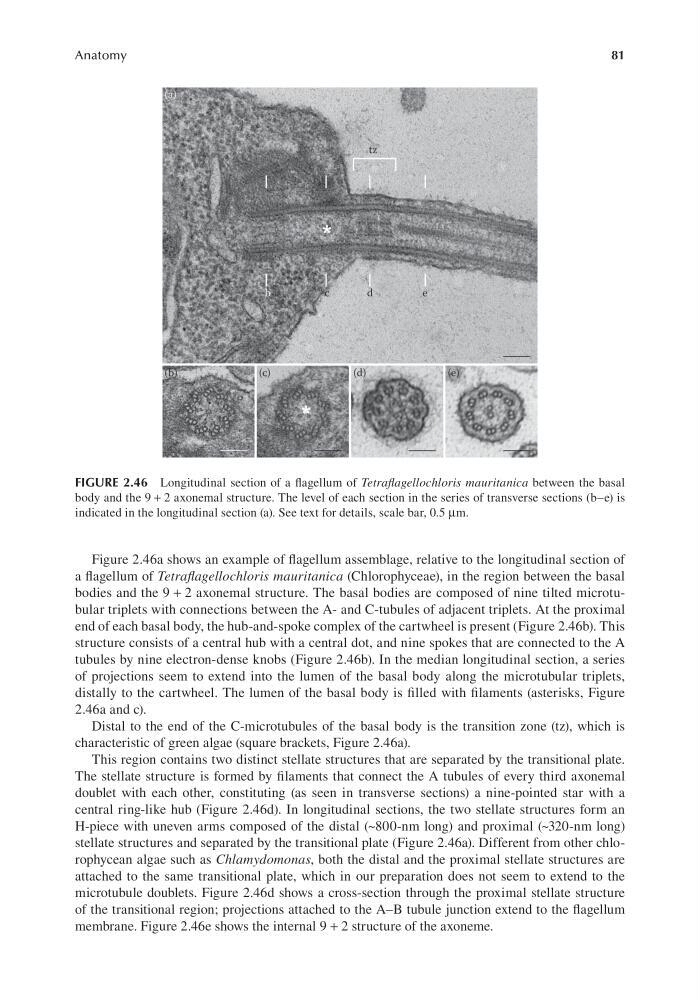
Figure 2.46a shows an example of flagellum assemblage, relative to the longitudinal section of
a flagellum of Tetraflagellochloris mauritanica (Chlorophyceae), in the region between the basal
bodies and the 9 + 2 axonemal structure. The basal bodies are composed of nine tilted microtubular triplets with connections between the A- and C-tubules of adjacent triplets. At the proximal
end of each basal body, the hub-and-spoke complex of the cartwheel is present (Figure 2.46b). This
structure consists of a central hub with a central dot, and nine spokes that are connected to the A
tubules by nine electron-dense knobs (Figure 2.46b). In the median longitudinal section, a series
of projections seem to extend into the lumen of the basal body along the microtubular triplets,
distally to the cartwheel. The lumen of the basal body is filled with filaments (asterisks, Figure
2.46a and c).
Distal to the end of the C-microtubules of the basal body is the transition zone (tz), which is
characteristic of green algae (square brackets, Figure 2.46a).
This region contains two distinct stellate structures that are separated by the transitional plate.
The stellate structure is formed by filaments that connect the A tubules of every third axonemal
doublet with each other, constituting (as seen in transverse sections) a nine-pointed star with a
central ring-like hub (Figure 2.46d). In longitudinal sections, the two stellate structures form an
H-piece with uneven arms composed of the distal (~800-nm long) and proximal (~320-nm long)
stellate structures and separated by the transitional plate (Figure 2.46a). Different from other chlorophycean algae such as Chlamydomonas, both the distal and the proximal stellate structures are
attached to the same transitional plate, which in our preparation does not seem to extend to the
microtubule doublets. Figure 2.46d shows a cross-section through the proximal stellate structure
of the transitional region; projections attached to the A–B tubule junction extend to the flagellum
membrane. Figure 2.46e shows the internal 9 + 2 structure of the axoneme.
FIGURE 2.46 Longitudinal section of a flagellum of Tetraflagellochloris mauritanica between the basal
body and the 9 + 2 axonemal structure. The level of each section in the series of transverse sections (b–e) is
indicated in the longitudinal section (a). See text for details, scale bar, 0.5 μm.
Anatomy
Figure 2.46a shows an example of flagellum assemblage, relative to the longitudinal section of
a flagellum of Tetraflagellochloris mauritanica (Chlorophyceae), in the region between the basal
bodies and the 9 + 2 axonemal structure. The basal bodies are composed of nine tilted microtubular triplets with connections between the A- and C-tubules of adjacent triplets. At the proximal
end of each basal body, the hub-and-spoke complex of the cartwheel is present (Figure 2.46b). This
structure consists of a central hub with a central dot, and nine spokes that are connected to the A
tubules by nine electron-dense knobs (Figure 2.46b). In the median longitudinal section, a series
of projections seem to extend into the lumen of the basal body along the microtubular triplets,
distally to the cartwheel. The lumen of the basal body is filled with filaments (asterisks, Figure
2.46a and c).
Distal to the end of the C-microtubules of the basal body is the transition zone (tz), which is
characteristic of green algae (square brackets, Figure 2.46a).
This region contains two distinct stellate structures that are separated by the transitional plate.
The stellate structure is formed by filaments that connect the A tubules of every third axonemal
doublet with each other, constituting (as seen in transverse sections) a nine-pointed star with a
central ring-like hub (Figure 2.46d). In longitudinal sections, the two stellate structures form an
H-piece with uneven arms composed of the distal (~800-nm long) and proximal (~320-nm long)
stellate structures and separated by the transitional plate (Figure 2.46a). Different from other chlorophycean algae such as Chlamydomonas, both the distal and the proximal stellate structures are
attached to the same transitional plate, which in our preparation does not seem to extend to the
microtubule doublets. Figure 2.46d shows a cross-section through the proximal stellate structure
of the transitional region; projections attached to the A–B tubule junction extend to the flagellum
membrane. Figure 2.46e shows the internal 9 + 2 structure of the axoneme.
FIGURE 2.46 Longitudinal section of a flagellum of Tetraflagellochloris mauritanica between the basal
body and the 9 + 2 axonemal structure. The level of each section in the series of transverse sections (b–e) is
indicated in the longitudinal section (a). See text for details, scale bar, 0.5 μm.