

52
Algae
Most taxa of the Prasinophytes (Chlorophyta) possess several scale types per cell, arranged in
1–5 layers on the surface of the cell body and flagella, those of each layer having a unique morphology for that taxon. These scales consist mainly of acidic polysaccharides involving unusual 2-keto
sugar acids, with glycoproteins as minor components. Members of the order Pyramimonadales
such as Pyramimonas sp. exhibit one of the most complex scaly covering among the Prasinophytes.
It consists of three layers of scales. The innermost scales are small, square, or pentagonal; the
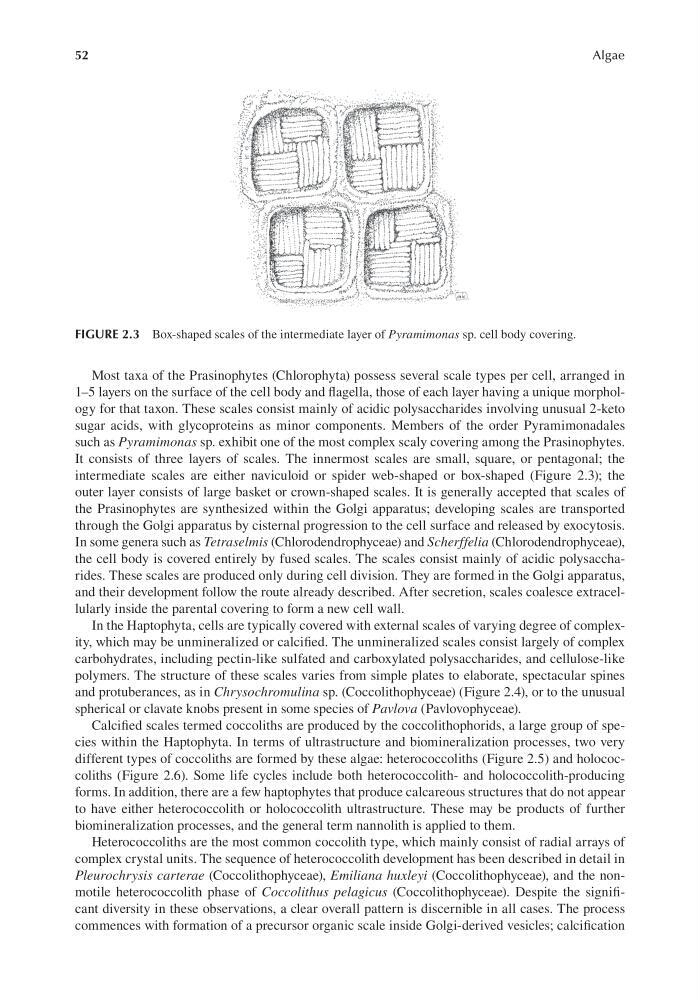
intermediate scales are either naviculoid or spider web-shaped or box-shaped (Figure 2.3); the
outer layer consists of large basket or crown-shaped scales. It is generally accepted that scales of
the Prasinophytes are synthesized within the Golgi apparatus; developing scales are transported
through the Golgi apparatus by cisternal progression to the cell surface and released by exocytosis.
In some genera such as Tetraselmis (Chlorodendrophyceae) and Scherffelia (Chlorodendrophyceae),
the cell body is covered entirely by fused scales. The scales consist mainly of acidic polysaccharides. These scales are produced only during cell division. They are formed in the Golgi apparatus,
and their development follow the route already described. After secretion, scales coalesce extracellularly inside the parental covering to form a new cell wall.
In the Haptophyta, cells are typically covered with external scales of varying degree of complexity, which may be unmineralized or calcified. The unmineralized scales consist largely of complex
carbohydrates, including pectin-like sulfated and carboxylated polysaccharides, and cellulose-like
polymers. The structure of these scales varies from simple plates to elaborate, spectacular spines
and protuberances, as in Chrysochromulina sp. (Coccolithophyceae) (Figure 2.4), or to the unusual
spherical or clavate knobs present in some species of Pavlova (Pavlovophyceae).
Calcified scales termed coccoliths are produced by the coccolithophorids, a large group of species within the Haptophyta. In terms of ultrastructure and biomineralization processes, two very
different types of coccoliths are formed by these algae: heterococcoliths (Figure 2.5) and holococcoliths (Figure 2.6). Some life cycles include both heterococcolith- and holococcolith-producing
forms. In addition, there are a few haptophytes that produce calcareous structures that do not appear
to have either heterococcolith or holococcolith ultrastructure. These may be products of further
biomineralization processes, and the general term nannolith is applied to them.
Heterococcoliths are the most common coccolith type, which mainly consist of radial arrays of
complex crystal units. The sequence of heterococcolith development has been described in detail in
Pleurochrysis carterae (Coccolithophyceae), Emiliana huxleyi (Coccolithophyceae), and the nonmotile heterococcolith phase of Coccolithus pelagicus (Coccolithophyceae). Despite the significant diversity in these observations, a clear overall pattern is discernible in all cases. The process
commences with formation of a precursor organic scale inside Golgi-derived vesicles; calcification
FIGURE 2.3 Box-shaped scales of the intermediate layer of Pyramimonas sp. cell body covering.
Algae
Most taxa of the Prasinophytes (Chlorophyta) possess several scale types per cell, arranged in
1–5 layers on the surface of the cell body and flagella, those of each layer having a unique morphology for that taxon. These scales consist mainly of acidic polysaccharides involving unusual 2-keto
sugar acids, with glycoproteins as minor components. Members of the order Pyramimonadales
such as Pyramimonas sp. exhibit one of the most complex scaly covering among the Prasinophytes.
It consists of three layers of scales. The innermost scales are small, square, or pentagonal; the
intermediate scales are either naviculoid or spider web-shaped or box-shaped (Figure 2.3); the
outer layer consists of large basket or crown-shaped scales. It is generally accepted that scales of
the Prasinophytes are synthesized within the Golgi apparatus; developing scales are transported
through the Golgi apparatus by cisternal progression to the cell surface and released by exocytosis.
In some genera such as Tetraselmis (Chlorodendrophyceae) and Scherffelia (Chlorodendrophyceae),
the cell body is covered entirely by fused scales. The scales consist mainly of acidic polysaccharides. These scales are produced only during cell division. They are formed in the Golgi apparatus,
and their development follow the route already described. After secretion, scales coalesce extracellularly inside the parental covering to form a new cell wall.
In the Haptophyta, cells are typically covered with external scales of varying degree of complexity, which may be unmineralized or calcified. The unmineralized scales consist largely of complex
carbohydrates, including pectin-like sulfated and carboxylated polysaccharides, and cellulose-like
polymers. The structure of these scales varies from simple plates to elaborate, spectacular spines
and protuberances, as in Chrysochromulina sp. (Coccolithophyceae) (Figure 2.4), or to the unusual
spherical or clavate knobs present in some species of Pavlova (Pavlovophyceae).
Calcified scales termed coccoliths are produced by the coccolithophorids, a large group of species within the Haptophyta. In terms of ultrastructure and biomineralization processes, two very
different types of coccoliths are formed by these algae: heterococcoliths (Figure 2.5) and holococcoliths (Figure 2.6). Some life cycles include both heterococcolith- and holococcolith-producing
forms. In addition, there are a few haptophytes that produce calcareous structures that do not appear
to have either heterococcolith or holococcolith ultrastructure. These may be products of further
biomineralization processes, and the general term nannolith is applied to them.
Heterococcoliths are the most common coccolith type, which mainly consist of radial arrays of
complex crystal units. The sequence of heterococcolith development has been described in detail in
Pleurochrysis carterae (Coccolithophyceae), Emiliana huxleyi (Coccolithophyceae), and the nonmotile heterococcolith phase of Coccolithus pelagicus (Coccolithophyceae). Despite the significant diversity in these observations, a clear overall pattern is discernible in all cases. The process
commences with formation of a precursor organic scale inside Golgi-derived vesicles; calcification
FIGURE 2.3 Box-shaped scales of the intermediate layer of Pyramimonas sp. cell body covering.