

23
General Overview
There are approximately 10 24 cyanobacterial cells in the oceans. To put that in perspective, the
number of cyanobacterial cells in the oceans is two orders of magnitude more than all the stars in
the sky. Pigmentation of cyanobacteria includes both chlorophyll a, blue and red phycobilins (phycoerythrin, phycocyanin, allophycocyanin), and carotenoids. These accessory pigments lie in the
phycobilisomes, located in rows on the outer surface of the thylakoids. Their thylakoids, which lie
free in the cytoplasm, are not arranged in stacks, but singled and equidistant, in contrast to prochlorophytes and most other algae, but similar to Rhodopyta and Glaucophyta.
The reserve polysaccharide is cyanophycean starch, stored in tiny granules lying between the
thylakoids. In addition, these cells often contain cyanophycin granules, that is, polymer of arginine
and aspartic acid. Some marine species also contain gas vesicles used for buoyancy regulation. In
some filamentous cyanobacteria, heterocysts and akinetes are formed. Heterocysts are vegetative
cells that have been drastically altered (loss of photosystem II, development of a thick, glycolipid
cell wall), to provide the necessary anoxygenic environment for the process of nitrogen fixation
(Figure 1.36). Some cyanobacteria produce potent hepato- and neurotoxins.
Prochlorophytes can be unicellular or filamentous, and depending on the filamentous species,
they can be either branched or unbranched. They exist as free-living components of pelagic nanoplankton and obligate symbionts within marine didemnid ascidians and holothurians and are mainly
limited to living in tropical and subtropical marine environments, with optimal growth temperature at about 24°C. Prochlorophytes possess chlorophyll a and b, as euglenoids and land plants, but
lack phycobilins, and this is the most significant difference between them and cyanobacteria, which
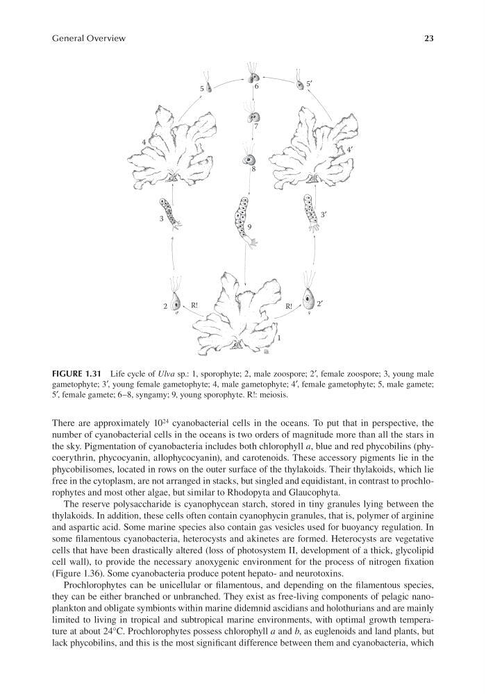
FIGURE 1.31 Life cycle of Ulva sp.: 1, sporophyte; 2, male zoospore; 2′, female zoospore; 3, young male
gametophyte; 3′, young female gametophyte; 4, male gametophyte; 4′, female gametophyte; 5, male gamete;
5′, female gamete; 6–8, syngamy; 9, young sporophyte. R!: meiosis.
General Overview
There are approximately 10 24 cyanobacterial cells in the oceans. To put that in perspective, the
number of cyanobacterial cells in the oceans is two orders of magnitude more than all the stars in
the sky. Pigmentation of cyanobacteria includes both chlorophyll a, blue and red phycobilins (phycoerythrin, phycocyanin, allophycocyanin), and carotenoids. These accessory pigments lie in the
phycobilisomes, located in rows on the outer surface of the thylakoids. Their thylakoids, which lie
free in the cytoplasm, are not arranged in stacks, but singled and equidistant, in contrast to prochlorophytes and most other algae, but similar to Rhodopyta and Glaucophyta.
The reserve polysaccharide is cyanophycean starch, stored in tiny granules lying between the
thylakoids. In addition, these cells often contain cyanophycin granules, that is, polymer of arginine
and aspartic acid. Some marine species also contain gas vesicles used for buoyancy regulation. In
some filamentous cyanobacteria, heterocysts and akinetes are formed. Heterocysts are vegetative
cells that have been drastically altered (loss of photosystem II, development of a thick, glycolipid
cell wall), to provide the necessary anoxygenic environment for the process of nitrogen fixation
(Figure 1.36). Some cyanobacteria produce potent hepato- and neurotoxins.
Prochlorophytes can be unicellular or filamentous, and depending on the filamentous species,
they can be either branched or unbranched. They exist as free-living components of pelagic nanoplankton and obligate symbionts within marine didemnid ascidians and holothurians and are mainly
limited to living in tropical and subtropical marine environments, with optimal growth temperature at about 24°C. Prochlorophytes possess chlorophyll a and b, as euglenoids and land plants, but
lack phycobilins, and this is the most significant difference between them and cyanobacteria, which
FIGURE 1.31 Life cycle of Ulva sp.: 1, sporophyte; 2, male zoospore; 2′, female zoospore; 3, young male
gametophyte; 3′, young female gametophyte; 4, male gametophyte; 4′, female gametophyte; 5, male gamete;
5′, female gamete; 6–8, syngamy; 9, young sporophyte. R!: meiosis.