

119
Anatomy
depending on the species. The amount of thylakoid membranes per cell is also a variable due to their
growth conditions and taxonomic specificity.
In most species, the thylakoids are arranged peripherally in 3–6 layers running parallel to the
cell membrane, forming an anastomosing network of concentric shells. This peripheral region has
been called chromatoplast, which is separated from the inner nucleoplasmic region called centroplasm. This type of thylakoid arrangement is characteristic of many unicellular and filamentous
cyanobacteria, such as Synechococeus planctibus, and Anabaena sp. In some other organisms such
as Oscillatoria and Arthrospira, the thylakoids are oriented perpendicular to the longitudinal cell
wall. Radial arrangement is present in Phormidium retzi. Thylakoids are not always restricted to the
cell periphery but can be found scattered throughout the cell as in Gloeotrichia sp. However, the
arrangement of thylakoids can change from cell to cell and cell type (vegetative cells, heterocyst,
akinete) within the same culture from parallel to a convoluted appearance. Thylakoids can fuse with
each other and form an anastomosing network; unlike the chloroplasts of eukaryotic algae, the thylakoids of cyanobacteria form stacking regions only to a very limited extent. During cell division,
the thylakoids have to be separated and divided into the daughter cells; generally, this division is an
active process. Cell division starts by a centripetal growth of cytoplasmic membrane and peptidoglycan layer, to form the septum; the thylakoid cylinder is narrowed at the level of the cross-wall by
invagination before the septum is formed. Proteins, lipids, carotenoids, and chlorophyll a are major
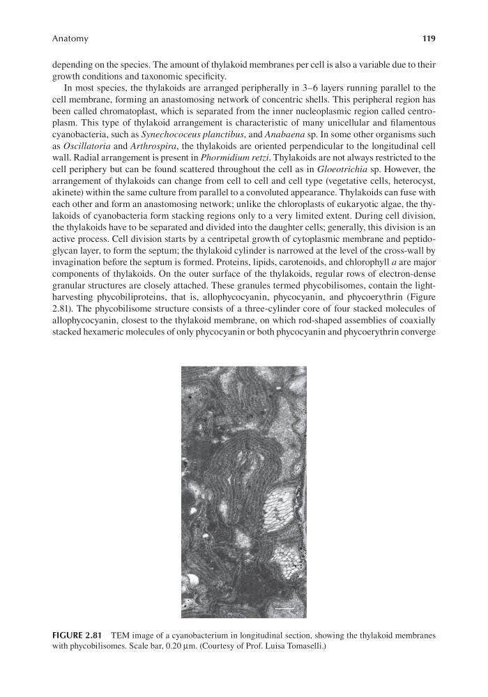
components of thylakoids. On the outer surface of the thylakoids, regular rows of electron-dense
granular structures are closely attached. These granules termed phycobilisomes, contain the lightharvesting phycobiliproteins, that is, allophycocyanin, phycocyanin, and phycoerythrin (Figure
2.81). The phycobilisome structure consists of a three-cylinder core of four stacked molecules of
allophycocyanin, closest to the thylakoid membrane, on which rod-shaped assemblies of coaxially
stacked hexameric molecules of only phycocyanin or both phycocyanin and phycoerythrin converge
FIGURE 2.81 TEM image of a cyanobacterium in longitudinal section, showing the thylakoid membranes
with phycobilisomes. Scale bar, 0.20 μm. (Courtesy of Prof. Luisa Tomaselli.)
Anatomy
depending on the species. The amount of thylakoid membranes per cell is also a variable due to their
growth conditions and taxonomic specificity.
In most species, the thylakoids are arranged peripherally in 3–6 layers running parallel to the
cell membrane, forming an anastomosing network of concentric shells. This peripheral region has
been called chromatoplast, which is separated from the inner nucleoplasmic region called centroplasm. This type of thylakoid arrangement is characteristic of many unicellular and filamentous
cyanobacteria, such as Synechococeus planctibus, and Anabaena sp. In some other organisms such
as Oscillatoria and Arthrospira, the thylakoids are oriented perpendicular to the longitudinal cell
wall. Radial arrangement is present in Phormidium retzi. Thylakoids are not always restricted to the
cell periphery but can be found scattered throughout the cell as in Gloeotrichia sp. However, the
arrangement of thylakoids can change from cell to cell and cell type (vegetative cells, heterocyst,
akinete) within the same culture from parallel to a convoluted appearance. Thylakoids can fuse with
each other and form an anastomosing network; unlike the chloroplasts of eukaryotic algae, the thylakoids of cyanobacteria form stacking regions only to a very limited extent. During cell division,
the thylakoids have to be separated and divided into the daughter cells; generally, this division is an
active process. Cell division starts by a centripetal growth of cytoplasmic membrane and peptidoglycan layer, to form the septum; the thylakoid cylinder is narrowed at the level of the cross-wall by
invagination before the septum is formed. Proteins, lipids, carotenoids, and chlorophyll a are major
components of thylakoids. On the outer surface of the thylakoids, regular rows of electron-dense
granular structures are closely attached. These granules termed phycobilisomes, contain the lightharvesting phycobiliproteins, that is, allophycocyanin, phycocyanin, and phycoerythrin (Figure
2.81). The phycobilisome structure consists of a three-cylinder core of four stacked molecules of
allophycocyanin, closest to the thylakoid membrane, on which rod-shaped assemblies of coaxially
stacked hexameric molecules of only phycocyanin or both phycocyanin and phycoerythrin converge
FIGURE 2.81 TEM image of a cyanobacterium in longitudinal section, showing the thylakoid membranes
with phycobilisomes. Scale bar, 0.20 μm. (Courtesy of Prof. Luisa Tomaselli.)