

112
Algae
Sampling Strategies
To determine the orientation of a stimulus field, an organism has to measure the stimulus intensity at
different positions, that is, it should detect either spatial or temporal patterns of light. Femtoplankton
(0.02–0.2 μm) is too small compared to the wavelength of light to create differential light intensity;
hence, it cannot determine the direction of a light source. Still, femtoplankton microorganisms can
use light, but can only measure its intensity and move in a light-intensity gradient. In contrast, phytoplankton dimensions are large enough to determine light direction and allow the scanning of the
environment by means of their directionally sensitive receptor.
Two fundamental and alternative strategies exist for obtaining information on light direction: parallel sampling and sequential sampling. In parallel sampling, the stimulus is detected by multiple
separated receptors positioned on different parts of the organism surface. In this case, the organism
measures directly the spatial gradient by simultaneous comparison of light intensities at two different parts of its body (one instant mechanism). This strategy is present in the zygote of the brown alga
Silvetia compressa; the photoreceptive system of the zygote is presumably the same as the egg before
fertilization. The zygote absorbs or scatters more than 95% of the effective blue wavelengths within
one cell diameter, thus creating a steep light gradient across the cells. Photoreceptors in or near the
plasma membrane are activated differentially, and the cellular response to this gradient of photoreceptor activation is to organize an axis and germinate from the darkest point. Photopolarization is an early
event, which has been observed 4 h after fertilization. The zygote produces a rhizoidal bulge at about
10 h after fertilization. Initially, any point on the cell surface is capable of becoming the germination
site. The bulge elongates by tip growth (i.e., addition of new cell membrane and wall material by
localized vesicle secretion), and about 18 h after fertilization the first cell division occurs. The plane
of division is perpendicular to the growth axis, resulting in the formation of two highly asymmetrical
cells with different developmental fates. The cell bearing the bulge will form the rhizoid, which will
show negative phototaxis, while the other cell will form the thallus. Figure 2.76 shows the first steps
of development of a Silvetia zygote from fertilization to rhizoid formation: changing of light direction
causes a change in the rhizoid growth axis.
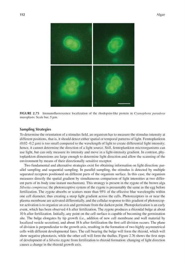
FIGURE 2.75 Immunofluorescence localization of the rhodopsin-like protein in Cyanophora paradoxa
muroplasts. Scale bar, 5 μm.
Algae
Sampling Strategies
To determine the orientation of a stimulus field, an organism has to measure the stimulus intensity at
different positions, that is, it should detect either spatial or temporal patterns of light. Femtoplankton
(0.02–0.2 μm) is too small compared to the wavelength of light to create differential light intensity;
hence, it cannot determine the direction of a light source. Still, femtoplankton microorganisms can
use light, but can only measure its intensity and move in a light-intensity gradient. In contrast, phytoplankton dimensions are large enough to determine light direction and allow the scanning of the
environment by means of their directionally sensitive receptor.
Two fundamental and alternative strategies exist for obtaining information on light direction: parallel sampling and sequential sampling. In parallel sampling, the stimulus is detected by multiple
separated receptors positioned on different parts of the organism surface. In this case, the organism
measures directly the spatial gradient by simultaneous comparison of light intensities at two different parts of its body (one instant mechanism). This strategy is present in the zygote of the brown alga
Silvetia compressa; the photoreceptive system of the zygote is presumably the same as the egg before
fertilization. The zygote absorbs or scatters more than 95% of the effective blue wavelengths within
one cell diameter, thus creating a steep light gradient across the cells. Photoreceptors in or near the
plasma membrane are activated differentially, and the cellular response to this gradient of photoreceptor activation is to organize an axis and germinate from the darkest point. Photopolarization is an early
event, which has been observed 4 h after fertilization. The zygote produces a rhizoidal bulge at about
10 h after fertilization. Initially, any point on the cell surface is capable of becoming the germination
site. The bulge elongates by tip growth (i.e., addition of new cell membrane and wall material by
localized vesicle secretion), and about 18 h after fertilization the first cell division occurs. The plane
of division is perpendicular to the growth axis, resulting in the formation of two highly asymmetrical
cells with different developmental fates. The cell bearing the bulge will form the rhizoid, which will
show negative phototaxis, while the other cell will form the thallus. Figure 2.76 shows the first steps
of development of a Silvetia zygote from fertilization to rhizoid formation: changing of light direction
causes a change in the rhizoid growth axis.
FIGURE 2.75 Immunofluorescence localization of the rhodopsin-like protein in Cyanophora paradoxa
muroplasts. Scale bar, 5 μm.