

θ helix ¼ cos
À1
ffiffiffiffiffiffiffiffiffiffiffiffiffiffiffiffiffiffiffiffi
2S helix þ 1
3
r
ð3:3Þ
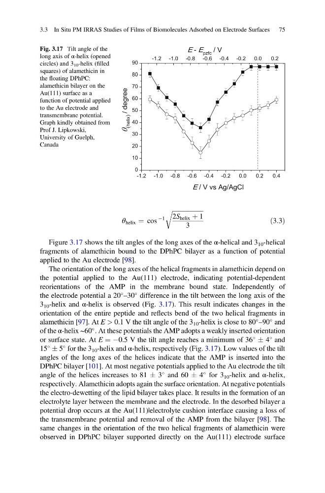
Figure 3.17 shows the tilt angles of the long axes of the α-helical and 3 10 -helical
fragments of alamethicin bound to the DPhPC bilayer as a function of potential
applied to the Au electrode [98].
The orientation of the long axes of the helical fragments in alamethicin depend on
the potential applied to the Au(111) electrode, indicating potential-dependent
reorientations of the AMP in the membrane bound state. Independently of
the electrode potential a 20
–30
difference in the tilt between the long axis of the
3 10 -helix and α-helix is observed (Fig. 3.17). This result indicates changes in the
orientation of the entire peptide and reflects bend of the two helical fragments in
alamethicin [97]. At E > 0.1 V the tilt angle of the 3 10 -helix is close to 80
–90
and
of the α-helix ~60
. At these potentials the AMP adopts a weakly inserted orientation
or surface state. At E ¼ À0.5 V the tilt angle reaches a minimum of 36
Æ 4
and
15
Æ 5
for the 3 10 -helix and α-helix, respectively (Fig. 3.17). Low values of the tilt
angles of the long axes of the helices indicate that the AMP is inserted into the
DPhPC bilayer [101]. At most negative potentials applied to the Au electrode the tilt
angle of the helices increases to 81 Æ 3
and 60 Æ 4
for 3 10 -helix and α-helix,
respectively. Alamethicin adopts again the surface orientation. At negative potentials
the electro-dewetting of the lipid bilayer takes place. It results in the formation of an
electrolyte layer between the membrane and the electrode. In the desorbed bilayer a
potential drop occurs at the Au(111)|electrolyte cushion interface causing a loss of
the transmembrane potential and removal of the AMP from the bilayer [98]. The
same changes in the orientation of the two helical fragments of alamethicin were
observed in DPhPC bilayer supported directly on the Au(111) electrode surface
-1.2 -1.0 -0.8 -0.6 -0.4 -0.2
0.0
0.2
0.4
0
10
20
30
40
50
60
70
80
90
E / V vs Ag/AgCl
(helix)
e
e
r
g
e
d
/
-1.2 -1.0 -0.8 -0.6 -0.4 -0.2
0.0
0.2
E - E pzfc / V
Fig. 3.17 Tilt angle of the
long axis of α-helix (opened
circles) and 3 10 -helix (filled
squares) of alamethicin in
the floating DPhPC:
alamethicin bilayer on the
Au(111) surface as a
function of potential applied
to the Au electrode and
transmembrane potential.
Graph kindly obtained from
Prof J. Lipkowski,
University of Guelph,
Canada
3.3 In Situ PM IRRAS Studies of Films of Biomolecules Adsorbed on Electrode Surfaces 75
À1
ffiffiffiffiffiffiffiffiffiffiffiffiffiffiffiffiffiffiffiffi
2S helix þ 1
3
r
ð3:3Þ
Figure 3.17 shows the tilt angles of the long axes of the α-helical and 3 10 -helical
fragments of alamethicin bound to the DPhPC bilayer as a function of potential
applied to the Au electrode [98].
The orientation of the long axes of the helical fragments in alamethicin depend on
the potential applied to the Au(111) electrode, indicating potential-dependent
reorientations of the AMP in the membrane bound state. Independently of
the electrode potential a 20
–30
difference in the tilt between the long axis of the
3 10 -helix and α-helix is observed (Fig. 3.17). This result indicates changes in the
orientation of the entire peptide and reflects bend of the two helical fragments in
alamethicin [97]. At E > 0.1 V the tilt angle of the 3 10 -helix is close to 80
–90
and
of the α-helix ~60
. At these potentials the AMP adopts a weakly inserted orientation
or surface state. At E ¼ À0.5 V the tilt angle reaches a minimum of 36
Æ 4
and
15
Æ 5
for the 3 10 -helix and α-helix, respectively (Fig. 3.17). Low values of the tilt
angles of the long axes of the helices indicate that the AMP is inserted into the
DPhPC bilayer [101]. At most negative potentials applied to the Au electrode the tilt
angle of the helices increases to 81 Æ 3
and 60 Æ 4
for 3 10 -helix and α-helix,
respectively. Alamethicin adopts again the surface orientation. At negative potentials
the electro-dewetting of the lipid bilayer takes place. It results in the formation of an
electrolyte layer between the membrane and the electrode. In the desorbed bilayer a
potential drop occurs at the Au(111)|electrolyte cushion interface causing a loss of
the transmembrane potential and removal of the AMP from the bilayer [98]. The
same changes in the orientation of the two helical fragments of alamethicin were
observed in DPhPC bilayer supported directly on the Au(111) electrode surface
-1.2 -1.0 -0.8 -0.6 -0.4 -0.2
0.0
0.2
0.4
0
10
20
30
40
50
60
70
80
90
E / V vs Ag/AgCl
(helix)
e
e
r
g
e
d
/
-1.2 -1.0 -0.8 -0.6 -0.4 -0.2
0.0
0.2
E - E pzfc / V
Fig. 3.17 Tilt angle of the
long axis of α-helix (opened
circles) and 3 10 -helix (filled
squares) of alamethicin in
the floating DPhPC:
alamethicin bilayer on the
Au(111) surface as a
function of potential applied
to the Au electrode and
transmembrane potential.
Graph kindly obtained from
Prof J. Lipkowski,
University of Guelph,
Canada
3.3 In Situ PM IRRAS Studies of Films of Biomolecules Adsorbed on Electrode Surfaces 75