

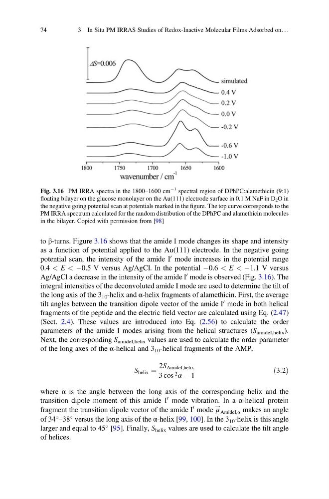
to β-turns. Figure 3.16 shows that the amide I mode changes its shape and intensity
as a function of potential applied to the Au(111) electrode. In the negative going
potential scan, the intensity of the amide I
0 mode increases in the potential range
0.4 < E < À0.5 V versus Ag/AgCl. In the potential À0.6 < E < À1.1 V versus
Ag/AgCl a decrease in the intensity of the amide I
0 mode is observed (Fig. 3.16). The
integral intensities of the deconvoluted amide I mode are used to determine the tilt of
the long axis of the 3 10 -helix and α-helix fragments of alamethicin. First, the average
tilt angles between the transition dipole vector of the amide I
0 mode in both helical
fragments of the peptide and the electric field vector are calculated using Eq. (2.47)
(Sect. 2.4). These values are introduced into Eq. (2.56) to calculate the order
parameters of the amide I modes arising from the helical structures (S amideI,helix ).
Next, the corresponding S amideI,helix values are used to calculate the order parameter
of the long axes of the α-helical and 3 10 -helical fragments of the AMP,
S helix ¼
2S AmideI,helix
3 cos 2 α À 1
ð3:2Þ
where α is the angle between the long axis of the corresponding helix and the
transition dipole moment of this amide I
0 mode vibration. In a α-helical protein
fragment the transition dipole vector of the amide I
0 mode μ
!
AmideI,α makes an angle
of 34
–38
versus the long axis of the α-helix [99, 100]. In the 3 10 -helix is this angle
larger and equal to 45
[95]. Finally, S helix values are used to calculate the tilt angle
of helices.
Fig. 3.16 PM IRRA spectra in the 1800–1600 cm
À1 spectral region of DPhPC:alamethicin (9:1)
floating bilayer on the glucose monolayer on the Au(111) electrode surface in 0.1 M NaF in D 2 O in
the negative going potential scan at potentials marked in the figure. The top curve corresponds to the
PM IRRA spectrum calculated for the random distribution of the DPhPC and alamethicin molecules
in the bilayer. Copied with permission from [98]
74
3 In Situ PM IRRAS Studies of Redox-Inactive Molecular Films Adsorbed on. . .
as a function of potential applied to the Au(111) electrode. In the negative going
potential scan, the intensity of the amide I
0 mode increases in the potential range
0.4 < E < À0.5 V versus Ag/AgCl. In the potential À0.6 < E < À1.1 V versus
Ag/AgCl a decrease in the intensity of the amide I
0 mode is observed (Fig. 3.16). The
integral intensities of the deconvoluted amide I mode are used to determine the tilt of
the long axis of the 3 10 -helix and α-helix fragments of alamethicin. First, the average
tilt angles between the transition dipole vector of the amide I
0 mode in both helical
fragments of the peptide and the electric field vector are calculated using Eq. (2.47)
(Sect. 2.4). These values are introduced into Eq. (2.56) to calculate the order
parameters of the amide I modes arising from the helical structures (S amideI,helix ).
Next, the corresponding S amideI,helix values are used to calculate the order parameter
of the long axes of the α-helical and 3 10 -helical fragments of the AMP,
S helix ¼
2S AmideI,helix
3 cos 2 α À 1
ð3:2Þ
where α is the angle between the long axis of the corresponding helix and the
transition dipole moment of this amide I
0 mode vibration. In a α-helical protein
fragment the transition dipole vector of the amide I
0 mode μ
!
AmideI,α makes an angle
of 34
–38
versus the long axis of the α-helix [99, 100]. In the 3 10 -helix is this angle
larger and equal to 45
[95]. Finally, S helix values are used to calculate the tilt angle
of helices.
Fig. 3.16 PM IRRA spectra in the 1800–1600 cm
À1 spectral region of DPhPC:alamethicin (9:1)
floating bilayer on the glucose monolayer on the Au(111) electrode surface in 0.1 M NaF in D 2 O in
the negative going potential scan at potentials marked in the figure. The top curve corresponds to the
PM IRRA spectrum calculated for the random distribution of the DPhPC and alamethicin molecules
in the bilayer. Copied with permission from [98]
74
3 In Situ PM IRRAS Studies of Redox-Inactive Molecular Films Adsorbed on. . .