

contribute to the secondary structure elements of the protein. Some fragments of the
polypeptide chain in a protein lack any ordered structure. These fragments are called
random coils. Various functional groups in the polypeptide chain absorb the IR light.
The amide I mode is the most important analytical band in the IR spectrum of
proteins, because it contains information about the secondary structure of the
protein. This band arises predominantly from the C¼O stretching vibration with a
small contribution from the CN stretching, CCN deformation and NH in plane
bending vibrations [85–87]. The position of the amide I mode in proteins is to a
small extent affected by the deuteration of the NH group, because of the negligible
contribution of the NH group in the entire amide I mode. The amide I mode of
proteins is usually broad, asymmetric, and composed of few superimposed bands
which arise from different secondary structure elements. In order to solve the
secondary structure of a protein two deconvolution procedures are used in the
literature:
1. Deconvolution of the amide I band after identifying the number and positions of
individual components from the second derivative of the IR spectrum or Fourierself-deconvolution spectra [86, 88, 89]
2. Decomposition of the amide I band into basis spectra which were calculated for a
calibration set of spectra of a protein with known structure [90–93]
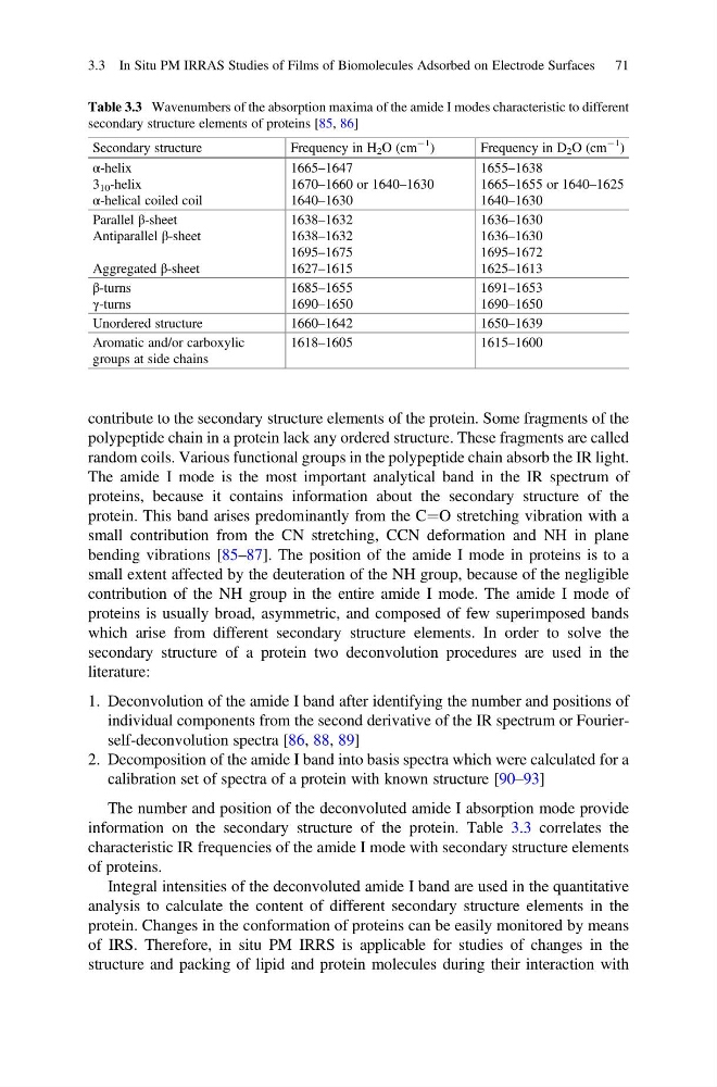
The number and position of the deconvoluted amide I absorption mode provide
information on the secondary structure of the protein. Table 3.3 correlates the
characteristic IR frequencies of the amide I mode with secondary structure elements
of proteins.
Integral intensities of the deconvoluted amide I band are used in the quantitative
analysis to calculate the content of different secondary structure elements in the
protein. Changes in the conformation of proteins can be easily monitored by means
of IRS. Therefore, in situ PM IRRS is applicable for studies of changes in the
structure and packing of lipid and protein molecules during their interaction with
Table 3.3 Wavenumbers of the absorption maxima of the amide I modes characteristic to different
secondary structure elements of proteins [85, 86]
Secondary structure
Frequency in H 2 O (cm
À1
)
Frequency in D 2 O (cm
À1
)
α-helix
3 10 -helix
α-helical coiled coil
1665–1647
1670–1660 or 1640–1630
1640–1630
1655–1638
1665–1655 or 1640–1625
1640–1630
Parallel β-sheet
Antiparallel β-sheet
Aggregated β-sheet
1638–1632
1638–1632
1695–1675
1627–1615
1636–1630
1636–1630
1695–1672
1625–1613
β-turns
γ-turns
1685–1655
1690–1650
1691–1653
1690–1650
Unordered structure
1660–1642
1650–1639
Aromatic and/or carboxylic
groups at side chains
1618–1605
1615–1600
3.3 In Situ PM IRRAS Studies of Films of Biomolecules Adsorbed on Electrode Surfaces 71
polypeptide chain in a protein lack any ordered structure. These fragments are called
random coils. Various functional groups in the polypeptide chain absorb the IR light.
The amide I mode is the most important analytical band in the IR spectrum of
proteins, because it contains information about the secondary structure of the
protein. This band arises predominantly from the C¼O stretching vibration with a
small contribution from the CN stretching, CCN deformation and NH in plane
bending vibrations [85–87]. The position of the amide I mode in proteins is to a
small extent affected by the deuteration of the NH group, because of the negligible
contribution of the NH group in the entire amide I mode. The amide I mode of
proteins is usually broad, asymmetric, and composed of few superimposed bands
which arise from different secondary structure elements. In order to solve the
secondary structure of a protein two deconvolution procedures are used in the
literature:
1. Deconvolution of the amide I band after identifying the number and positions of
individual components from the second derivative of the IR spectrum or Fourierself-deconvolution spectra [86, 88, 89]
2. Decomposition of the amide I band into basis spectra which were calculated for a
calibration set of spectra of a protein with known structure [90–93]
The number and position of the deconvoluted amide I absorption mode provide
information on the secondary structure of the protein. Table 3.3 correlates the
characteristic IR frequencies of the amide I mode with secondary structure elements
of proteins.
Integral intensities of the deconvoluted amide I band are used in the quantitative
analysis to calculate the content of different secondary structure elements in the
protein. Changes in the conformation of proteins can be easily monitored by means
of IRS. Therefore, in situ PM IRRS is applicable for studies of changes in the
structure and packing of lipid and protein molecules during their interaction with
Table 3.3 Wavenumbers of the absorption maxima of the amide I modes characteristic to different
secondary structure elements of proteins [85, 86]
Secondary structure
Frequency in H 2 O (cm
À1
)
Frequency in D 2 O (cm
À1
)
α-helix
3 10 -helix
α-helical coiled coil
1665–1647
1670–1660 or 1640–1630
1640–1630
1655–1638
1665–1655 or 1640–1625
1640–1630
Parallel β-sheet
Antiparallel β-sheet
Aggregated β-sheet
1638–1632
1638–1632
1695–1675
1627–1615
1636–1630
1636–1630
1695–1672
1625–1613
β-turns
γ-turns
1685–1655
1690–1650
1691–1653
1690–1650
Unordered structure
1660–1642
1650–1639
Aromatic and/or carboxylic
groups at side chains
1618–1605
1615–1600
3.3 In Situ PM IRRAS Studies of Films of Biomolecules Adsorbed on Electrode Surfaces 71