

The procedure of the fabrication of floating lipid bilayers on the Au electrode
surface was introduced by Kycia et al. [33]. It is an alternative method to the
fabrication of supported lipid bilayers. Figure 3.12 shows schematically the structure
and composition of a floating lipid bilayer.
First, 1-thio-β-D-glucose is self-assembled on the gold surface. This modification
ensures the hydrophilic environment for the inner leaflet of the model membrane
[33]. Next, LB-LS transfers are used to prepare the inner leaflet and outer leaflets of
the lipid membrane. In a floating lipid bilayer both sides of the membrane are in
direct contact with aqueous environment, representing an excellent model for the
incorporation of transmembrane proteins [33]. A floating DMPC lipid bilayer was
fabricated on thioglucose monolayer surface, which was self-assembled on the gold
surface. The capacitances of the supported and floating lipid DMPC bilayers are
similar. The capacitance minimum of the floating DMPC bilayer is equal to
7 μF cm
À2 [78]. In the negative going potential scan an increase in the capacitance
is observed (at E < À0.40 V vs Ag/AgCl). The bilayer desorbs from the Au surface
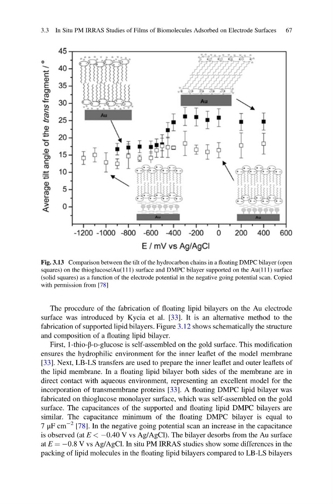
at E ¼ À0.8 V vs Ag/AgCl. In situ PM IRRAS studies show some differences in the
packing of lipid molecules in the floating lipid bilayers compared to LB-LS bilayers
Fig. 3.13 Comparison between the tilt of the hydrocarbon chains in a floating DMPC bilayer (open
squares) on the thioglucose|Au(111) surface and DMPC bilayer supported on the Au(111) surface
(solid squares) as a function of the electrode potential in the negative going potential scan. Copied
with permission from [78]
3.3 In Situ PM IRRAS Studies of Films of Biomolecules Adsorbed on Electrode Surfaces 67
surface was introduced by Kycia et al. [33]. It is an alternative method to the
fabrication of supported lipid bilayers. Figure 3.12 shows schematically the structure
and composition of a floating lipid bilayer.
First, 1-thio-β-D-glucose is self-assembled on the gold surface. This modification
ensures the hydrophilic environment for the inner leaflet of the model membrane
[33]. Next, LB-LS transfers are used to prepare the inner leaflet and outer leaflets of
the lipid membrane. In a floating lipid bilayer both sides of the membrane are in
direct contact with aqueous environment, representing an excellent model for the
incorporation of transmembrane proteins [33]. A floating DMPC lipid bilayer was
fabricated on thioglucose monolayer surface, which was self-assembled on the gold
surface. The capacitances of the supported and floating lipid DMPC bilayers are
similar. The capacitance minimum of the floating DMPC bilayer is equal to
7 μF cm
À2 [78]. In the negative going potential scan an increase in the capacitance
is observed (at E < À0.40 V vs Ag/AgCl). The bilayer desorbs from the Au surface
at E ¼ À0.8 V vs Ag/AgCl. In situ PM IRRAS studies show some differences in the
packing of lipid molecules in the floating lipid bilayers compared to LB-LS bilayers
Fig. 3.13 Comparison between the tilt of the hydrocarbon chains in a floating DMPC bilayer (open
squares) on the thioglucose|Au(111) surface and DMPC bilayer supported on the Au(111) surface
(solid squares) as a function of the electrode potential in the negative going potential scan. Copied
with permission from [78]
3.3 In Situ PM IRRAS Studies of Films of Biomolecules Adsorbed on Electrode Surfaces 67