

oriented toward the hydrocarbon chain region of the inner leaflet. Deconvolution of
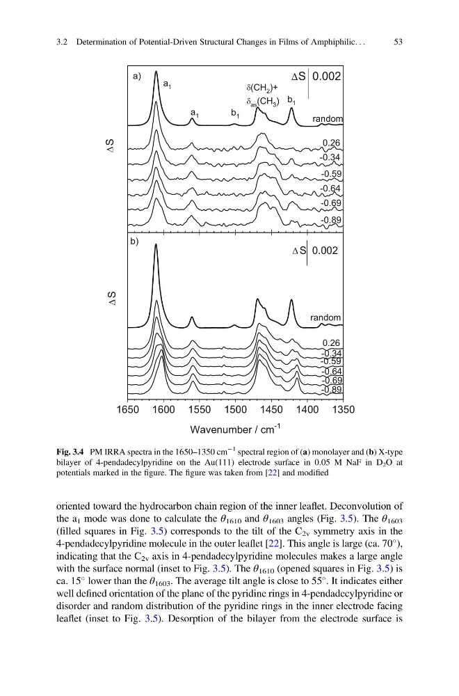
the a 1 mode was done to calculate the θ 1610 and θ 1603 angles (Fig. 3.5). The θ 1603
(filled squares in Fig. 3.5) corresponds to the tilt of the C 2v symmetry axis in the
4-pendadecylpyridine molecule in the outer leaflet [22]. This angle is large (ca. 70
),
indicating that the C 2v axis in 4-pendadecylpyridine molecules makes a large angle
with the surface normal (inset to Fig. 3.5). The θ 1610 (opened squares in Fig. 3.5) is
ca. 15
lower than the θ 1603 . The average tilt angle is close to 55
. It indicates either
well defined orientation of the plane of the pyridine rings in 4-pendadecylpyridine or
disorder and random distribution of the pyridine rings in the inner electrode facing
leaflet (inset to Fig. 3.5). Desorption of the bilayer from the electrode surface is
1650 1600 1550 1500 1450 1400 1350
random
0.002
b)
S
Wavenumber / cm
-1
-0.89
-0.69
-0.64
-0.59
0.26
-0.34
b 1
a 1
b 1
-0.89
-0.69
-0.64
-0.59
S
S 0.002
a)
random
0.26
-0.34
a 1
(CH 2 )+
as (CH 3 )
S
Fig. 3.4 PM IRRA spectra in the 1650–1350 cm
À1 spectral region of (a) monolayer and (b) X-type
bilayer of 4-pendadecylpyridine on the Au(111) electrode surface in 0.05 M NaF in D 2 O at
potentials marked in the figure. The figure was taken from [22] and modified
3.2 Determination of Potential-Driven Structural Changes in Films of Amphiphilic. . .
53
the a 1 mode was done to calculate the θ 1610 and θ 1603 angles (Fig. 3.5). The θ 1603
(filled squares in Fig. 3.5) corresponds to the tilt of the C 2v symmetry axis in the
4-pendadecylpyridine molecule in the outer leaflet [22]. This angle is large (ca. 70
),
indicating that the C 2v axis in 4-pendadecylpyridine molecules makes a large angle
with the surface normal (inset to Fig. 3.5). The θ 1610 (opened squares in Fig. 3.5) is
ca. 15
lower than the θ 1603 . The average tilt angle is close to 55
. It indicates either
well defined orientation of the plane of the pyridine rings in 4-pendadecylpyridine or
disorder and random distribution of the pyridine rings in the inner electrode facing
leaflet (inset to Fig. 3.5). Desorption of the bilayer from the electrode surface is
1650 1600 1550 1500 1450 1400 1350
random
0.002
b)
S
Wavenumber / cm
-1
-0.89
-0.69
-0.64
-0.59
0.26
-0.34
b 1
a 1
b 1
-0.89
-0.69
-0.64
-0.59
S
S 0.002
a)
random
0.26
-0.34
a 1
(CH 2 )+
as (CH 3 )
S
Fig. 3.4 PM IRRA spectra in the 1650–1350 cm
À1 spectral region of (a) monolayer and (b) X-type
bilayer of 4-pendadecylpyridine on the Au(111) electrode surface in 0.05 M NaF in D 2 O at
potentials marked in the figure. The figure was taken from [22] and modified
3.2 Determination of Potential-Driven Structural Changes in Films of Amphiphilic. . .
53