

4.3 Washing Treatment for Bacterial Isolation from
Sclerotia Grains
Sclerotia grains collected from soil are in contact with soil, so bacteria derived from
soil are attached to the outer surface of the sclerotia grains. Because of the special
honeycomb-like structure of the sclerotia grains, it is necessary to consider a method
for collecting only the bacteria that live inside the sclerotium. Here we introduce the
treatment designed by Ohta et al. (2003) for the isolation of bacteria from sclerotia
grains.
The sclerotia grains used for testing this treatment were obtained from Tsubame
soil (Fulvic Andosols, WRB/ FAO-UNESCO) from Mt. Myoko, Niigata Prefecture,
Japan (36
54
0 00
00 N, 138
8
0 25
00 E, altitude: 1330 m). Sclerotia grains were carefully
picked from the soil and soil particles on the grain surface were removed using
tweezers. The washing procedure for bacterial isolation from sclerotia grains is
diagramed in Fig. 4.2. To remove surface-attached soil, a sclerotium grain was
placed in a micro-centrifuge tube containing 1 mL of sterile water and washed by
vortexing for 1 min, after which the wash solution was removed from the tube using
a sterile pipette. The number of viable bacteria in the wash solution was determined
by plate count. After ten repetitions of the washing treatment, the washed grain was
crushed with a sterile glass rod in 1 mL of sterile water and ultrasonicated for 2 min
at 100 W. The number of viable bacteria released by crushing and ultrasonication
was determined by plate count.
The effects of repeated washes and the final ultrasonication on plate counts of
bacteria from a sclerotium grain were then examined. The results of three independent experiments are shown in Fig. 4.3. The first sample (sample A; fresh weight not
determined) yielded a plate count of 3.05 Â 10
3 CFU in the first wash fraction. The
third and tenth wash fractions had relative plate counts that were only 3.6% and
0.7%, respectively, of the first wash fraction. The ultrasonication of the washed grain
yielded a plate count 7.9% of the plate count of the first wash fraction, suggesting a
large release of bacteria from the inside of the sclerotium. This was also the case for
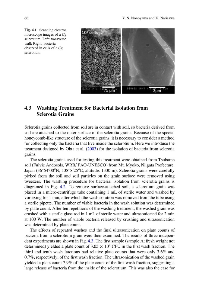
Fig. 4.1 Scanning electron
microscope images of a Cg
sclerotium. Left: transverse
wall; Right: bacteria
observed in cells of a Cg
sclerotium
66
Y. S. Nonoyama and K. Narisawa
Sclerotia Grains
Sclerotia grains collected from soil are in contact with soil, so bacteria derived from
soil are attached to the outer surface of the sclerotia grains. Because of the special
honeycomb-like structure of the sclerotia grains, it is necessary to consider a method
for collecting only the bacteria that live inside the sclerotium. Here we introduce the
treatment designed by Ohta et al. (2003) for the isolation of bacteria from sclerotia
grains.
The sclerotia grains used for testing this treatment were obtained from Tsubame
soil (Fulvic Andosols, WRB/ FAO-UNESCO) from Mt. Myoko, Niigata Prefecture,
Japan (36
54
0 00
00 N, 138
8
0 25
00 E, altitude: 1330 m). Sclerotia grains were carefully
picked from the soil and soil particles on the grain surface were removed using
tweezers. The washing procedure for bacterial isolation from sclerotia grains is
diagramed in Fig. 4.2. To remove surface-attached soil, a sclerotium grain was
placed in a micro-centrifuge tube containing 1 mL of sterile water and washed by
vortexing for 1 min, after which the wash solution was removed from the tube using
a sterile pipette. The number of viable bacteria in the wash solution was determined
by plate count. After ten repetitions of the washing treatment, the washed grain was
crushed with a sterile glass rod in 1 mL of sterile water and ultrasonicated for 2 min
at 100 W. The number of viable bacteria released by crushing and ultrasonication
was determined by plate count.
The effects of repeated washes and the final ultrasonication on plate counts of
bacteria from a sclerotium grain were then examined. The results of three independent experiments are shown in Fig. 4.3. The first sample (sample A; fresh weight not
determined) yielded a plate count of 3.05 Â 10
3 CFU in the first wash fraction. The
third and tenth wash fractions had relative plate counts that were only 3.6% and
0.7%, respectively, of the first wash fraction. The ultrasonication of the washed grain
yielded a plate count 7.9% of the plate count of the first wash fraction, suggesting a
large release of bacteria from the inside of the sclerotium. This was also the case for
Fig. 4.1 Scanning electron
microscope images of a Cg
sclerotium. Left: transverse
wall; Right: bacteria
observed in cells of a Cg
sclerotium
66
Y. S. Nonoyama and K. Narisawa