

sterilization and to preserve the mesofauna for further examination. Mesofauna were
identified to genus under a digital high-density video microscope (VH-7000,
Keyence, Osaka, Japan).
3.2.1.3 Isolation of Fungal DNA from Soil Mesofauna
Three to five individuals of each species from each subplot were crushed using a
metal crusher (TAITEC Corporation, Saitama, Japan) and fungal DNA was isolated
using Prepman™ Ultra sample reagent (Applied Biosystems, CA, USA). This
process was repeated three times for each location, and results were averaged. The
ribosomal DNA internal transcribed spacer (ITS) regions were then amplified by
PCR using the ITS-1F/LR21 primer pair and Taq DNA polymerase (Applied
Biosystems, Waltham, MA, USA) with conditions as follows: a hot start at 96
C
for 30 s, then 35 cycles consisting of 10 s at 96
C, 30 s at 55
C, and 30 s at 72
C.
Samples were also subjected to T-RFLP analysis. T-RFLP refers to the use of
fluorescently labeled primers combined with restriction digestion to visualize
sequence variation in either single- or mixed-species DNA samples (Dickie and
Fitz John 2007). The T-RFLP technique was first developed by Liu et al. (1997) as a
tool for assessing bacterial diversity and comparing bacterial community structure
between environmental samples (Marsh 1999; Lukow et al. 2000; Kitts 2001).
T-RFLP data are visualized as an electropherogram, with the size and relative
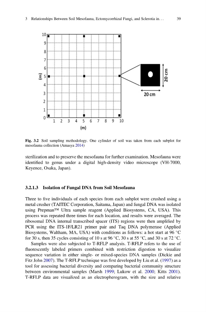
Fig. 3.2 Soil sampling methodology. One cylinder of soil was taken from each subplot for
mesofauna collection (Amasya 2014)
3 Relationships Between Soil Mesofauna, Ectomycorrhizal Fungi, and Sclerotia in. . .
39
identified to genus under a digital high-density video microscope (VH-7000,
Keyence, Osaka, Japan).
3.2.1.3 Isolation of Fungal DNA from Soil Mesofauna
Three to five individuals of each species from each subplot were crushed using a
metal crusher (TAITEC Corporation, Saitama, Japan) and fungal DNA was isolated
using Prepman™ Ultra sample reagent (Applied Biosystems, CA, USA). This
process was repeated three times for each location, and results were averaged. The
ribosomal DNA internal transcribed spacer (ITS) regions were then amplified by
PCR using the ITS-1F/LR21 primer pair and Taq DNA polymerase (Applied
Biosystems, Waltham, MA, USA) with conditions as follows: a hot start at 96
C
for 30 s, then 35 cycles consisting of 10 s at 96
C, 30 s at 55
C, and 30 s at 72
C.
Samples were also subjected to T-RFLP analysis. T-RFLP refers to the use of
fluorescently labeled primers combined with restriction digestion to visualize
sequence variation in either single- or mixed-species DNA samples (Dickie and
Fitz John 2007). The T-RFLP technique was first developed by Liu et al. (1997) as a
tool for assessing bacterial diversity and comparing bacterial community structure
between environmental samples (Marsh 1999; Lukow et al. 2000; Kitts 2001).
T-RFLP data are visualized as an electropherogram, with the size and relative
Fig. 3.2 Soil sampling methodology. One cylinder of soil was taken from each subplot for
mesofauna collection (Amasya 2014)
3 Relationships Between Soil Mesofauna, Ectomycorrhizal Fungi, and Sclerotia in. . .
39