

77
Pulsed Electric Fields in Biological Cells and Membranes
(∼5–7 nm); and C m the membrane capacitance, with ε m denoting the dielectric permittivity of the membrane. The transmembrane voltage could then be derived from
Equation 2.1 for an external alternating current field. In the frequency domain, the
transmembrane voltage ∆Φ could be expressed as follows (Grosse and Schwan 1992):
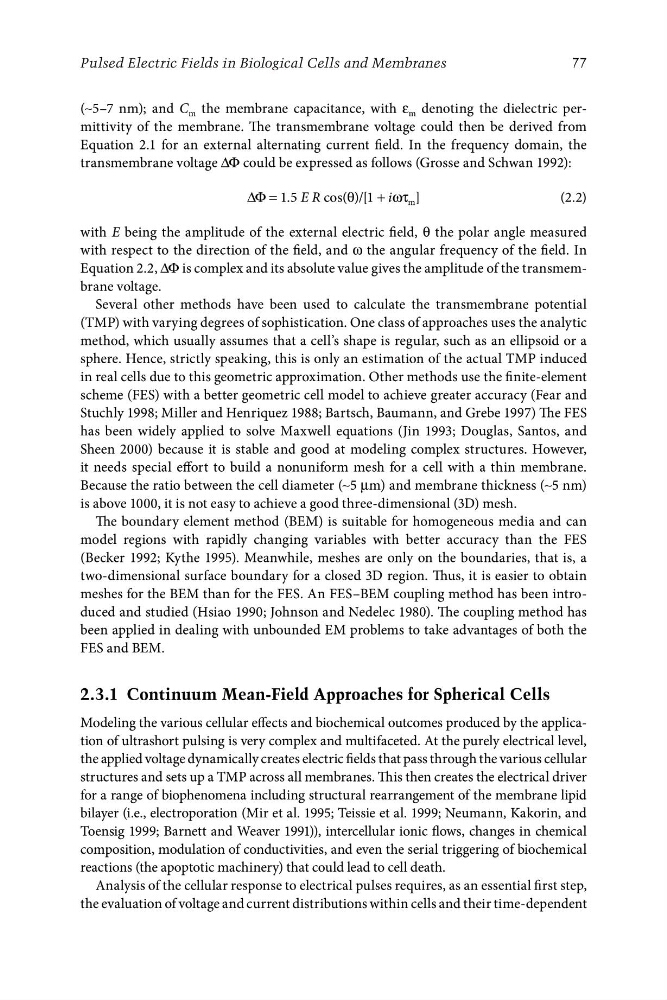
∆Φ = 1.5 E R cos(θ)/[1 + iωτ m ]
(2.2)
with E being the amplitude of the external electric field, θ the polar angle measured
with respect to the direction of the field, and ω the angular frequency of the field. In
Equation 2.2, ∆Φ is complex and its absolute value gives the amplitude of the transmembrane voltage.
Several other methods have been used to calculate the transmembrane potential
(TMP) with varying degrees of sophistication. One class of approaches uses the analytic
method, which usually assumes that a cell’s shape is regular, such as an ellipsoid or a
sphere. Hence, strictly speaking, this is only an estimation of the actual TMP induced
in real cells due to this geometric approximation. Other methods use the finite-element
scheme (FES) with a better geometric cell model to achieve greater accuracy (Fear and
Stuchly 1998; Miller and Henriquez 1988; Bartsch, Baumann, and Grebe 1997) The FES
has been widely applied to solve Maxwell equations (Jin 1993; Douglas, Santos, and
Sheen 2000) because it is stable and good at modeling complex structures. However,
it needs special effort to build a nonuniform mesh for a cell with a thin membrane.
Because the ratio between the cell diameter (∼5 μm) and membrane thickness (∼5 nm)
is above 1000, it is not easy to achieve a good three-dimensional (3D) mesh.
The boundary element method (BEM) is suitable for homogeneous media and can
model regions with rapidly changing variables with better accuracy than the FES
(Becker 1992; Kythe 1995). Meanwhile, meshes are only on the boundaries, that is, a
two-dimensional surface boundary for a closed 3D region. Thus, it is easier to obtain
meshes for the BEM than for the FES. An FES–BEM coupling method has been introduced and studied (Hsiao 1990; Johnson and Nedelec 1980). The coupling method has
been applied in dealing with unbounded EM problems to take advantages of both the
FES and BEM.
2.3.1 Continuum Mean-Field Approaches for Spherical Cells
Modeling the various cellular effects and biochemical outcomes produced by the application of ultrashort pulsing is very complex and multifaceted. At the purely electrical level,
the applied voltage dynamically creates electric fields that pass through the various cellular
structures and sets up a TMP across all membranes. This then creates the electrical driver
for a range of biophenomena including structural rearrangement of the membrane lipid
bilayer (i.e., electroporation (Mir et al. 1995; Teissie et al. 1999; Neumann, Kakorin, and
Toensig 1999; Barnett and Weaver 1991)), intercellular ionic flows, changes in chemical
composition, modulation of conductivities, and even the serial triggering of biochemical
reactions (the apoptotic machinery) that could lead to cell death.
Analysis of the cellular response to electrical pulses requires, as an essential first step,
the evaluation of voltage and current distributions within cells and their time-dependent
Pulsed Electric Fields in Biological Cells and Membranes
(∼5–7 nm); and C m the membrane capacitance, with ε m denoting the dielectric permittivity of the membrane. The transmembrane voltage could then be derived from
Equation 2.1 for an external alternating current field. In the frequency domain, the
transmembrane voltage ∆Φ could be expressed as follows (Grosse and Schwan 1992):
∆Φ = 1.5 E R cos(θ)/[1 + iωτ m ]
(2.2)
with E being the amplitude of the external electric field, θ the polar angle measured
with respect to the direction of the field, and ω the angular frequency of the field. In
Equation 2.2, ∆Φ is complex and its absolute value gives the amplitude of the transmembrane voltage.
Several other methods have been used to calculate the transmembrane potential
(TMP) with varying degrees of sophistication. One class of approaches uses the analytic
method, which usually assumes that a cell’s shape is regular, such as an ellipsoid or a
sphere. Hence, strictly speaking, this is only an estimation of the actual TMP induced
in real cells due to this geometric approximation. Other methods use the finite-element
scheme (FES) with a better geometric cell model to achieve greater accuracy (Fear and
Stuchly 1998; Miller and Henriquez 1988; Bartsch, Baumann, and Grebe 1997) The FES
has been widely applied to solve Maxwell equations (Jin 1993; Douglas, Santos, and
Sheen 2000) because it is stable and good at modeling complex structures. However,
it needs special effort to build a nonuniform mesh for a cell with a thin membrane.
Because the ratio between the cell diameter (∼5 μm) and membrane thickness (∼5 nm)
is above 1000, it is not easy to achieve a good three-dimensional (3D) mesh.
The boundary element method (BEM) is suitable for homogeneous media and can
model regions with rapidly changing variables with better accuracy than the FES
(Becker 1992; Kythe 1995). Meanwhile, meshes are only on the boundaries, that is, a
two-dimensional surface boundary for a closed 3D region. Thus, it is easier to obtain
meshes for the BEM than for the FES. An FES–BEM coupling method has been introduced and studied (Hsiao 1990; Johnson and Nedelec 1980). The coupling method has
been applied in dealing with unbounded EM problems to take advantages of both the
FES and BEM.
2.3.1 Continuum Mean-Field Approaches for Spherical Cells
Modeling the various cellular effects and biochemical outcomes produced by the application of ultrashort pulsing is very complex and multifaceted. At the purely electrical level,
the applied voltage dynamically creates electric fields that pass through the various cellular
structures and sets up a TMP across all membranes. This then creates the electrical driver
for a range of biophenomena including structural rearrangement of the membrane lipid
bilayer (i.e., electroporation (Mir et al. 1995; Teissie et al. 1999; Neumann, Kakorin, and
Toensig 1999; Barnett and Weaver 1991)), intercellular ionic flows, changes in chemical
composition, modulation of conductivities, and even the serial triggering of biochemical
reactions (the apoptotic machinery) that could lead to cell death.
Analysis of the cellular response to electrical pulses requires, as an essential first step,
the evaluation of voltage and current distributions within cells and their time-dependent