

76
Electromagnetic Fields in Biological Systems
A B
C
D V E = 0V
H
F G
I
σ > 0
σ > 0
σ = 0
σ = 0
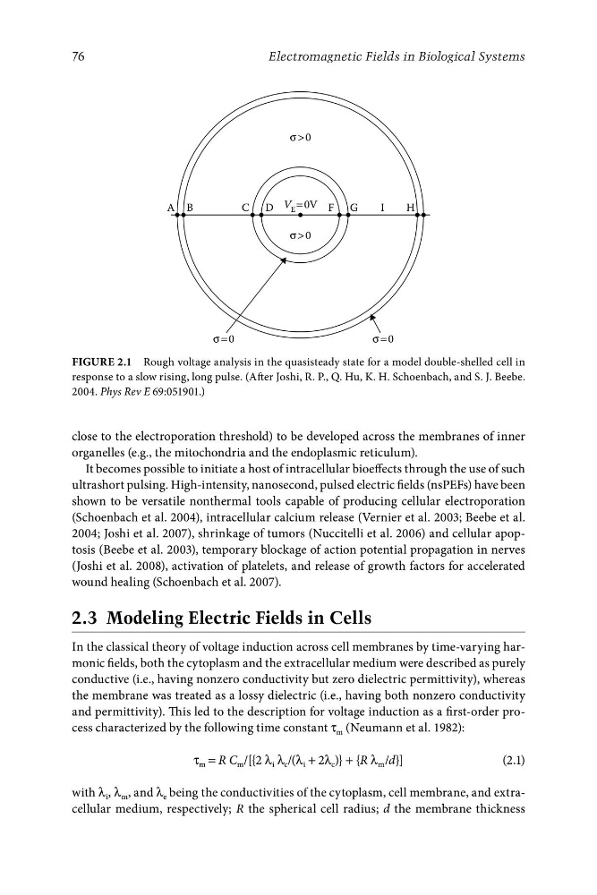
Figure 2.1 Rough voltage analysis in the quasisteady state for a model double-shelled cell in
response to a slow rising, long pulse. (After Joshi, R. P., Q. Hu, K. H. Schoenbach, and S. J. Beebe.
2004. Phys Rev E 69:051901.)
close to the electroporation threshold) to be developed across the membranes of inner
organelles (e.g., the mitochondria and the endoplasmic reticulum).
It becomes possible to initiate a host of intracellular bioeffects through the use of such
ultrashort pulsing. High-intensity, nanosecond, pulsed electric fields (nsPEFs) have been
shown to be versatile nonthermal tools capable of producing cellular electroporation
(Schoenbach et al. 2004), intracellular calcium release (Vernier et al. 2003; Beebe et al.
2004; Joshi et al. 2007), shrinkage of tumors (Nuccitelli et al. 2006) and cellular apoptosis (Beebe et al. 2003), temporary blockage of action potential propagation in nerves
(Joshi et al. 2008), activation of platelets, and release of growth factors for accelerated
wound healing (Schoenbach et al. 2007).
2.3 Modeling Electric Fields in Cells
In the classical theory of voltage induction across cell membranes by time-varying harmonic fields, both the cytoplasm and the extracellular medium were described as purely
conductive (i.e., having nonzero conductivity but zero dielectric permittivity), whereas
the membrane was treated as a lossy dielectric (i.e., having both nonzero conductivity
and permittivity). This led to the description for voltage induction as a first-order process characterized by the following time constant τ m (Neumann et al. 1982):
τ m = R C m /[{2 λ i λ c /(λ i + 2λ c )} + {R λ m /d}]
(2.1)
with λ i , λ m , and λ e being the conductivities of the cytoplasm, cell membrane, and extracellular medium, respectively; R the spherical cell radius; d the membrane thickness
Electromagnetic Fields in Biological Systems
A B
C
D V E = 0V
H
F G
I
σ > 0
σ > 0
σ = 0
σ = 0
Figure 2.1 Rough voltage analysis in the quasisteady state for a model double-shelled cell in
response to a slow rising, long pulse. (After Joshi, R. P., Q. Hu, K. H. Schoenbach, and S. J. Beebe.
2004. Phys Rev E 69:051901.)
close to the electroporation threshold) to be developed across the membranes of inner
organelles (e.g., the mitochondria and the endoplasmic reticulum).
It becomes possible to initiate a host of intracellular bioeffects through the use of such
ultrashort pulsing. High-intensity, nanosecond, pulsed electric fields (nsPEFs) have been
shown to be versatile nonthermal tools capable of producing cellular electroporation
(Schoenbach et al. 2004), intracellular calcium release (Vernier et al. 2003; Beebe et al.
2004; Joshi et al. 2007), shrinkage of tumors (Nuccitelli et al. 2006) and cellular apoptosis (Beebe et al. 2003), temporary blockage of action potential propagation in nerves
(Joshi et al. 2008), activation of platelets, and release of growth factors for accelerated
wound healing (Schoenbach et al. 2007).
2.3 Modeling Electric Fields in Cells
In the classical theory of voltage induction across cell membranes by time-varying harmonic fields, both the cytoplasm and the extracellular medium were described as purely
conductive (i.e., having nonzero conductivity but zero dielectric permittivity), whereas
the membrane was treated as a lossy dielectric (i.e., having both nonzero conductivity
and permittivity). This led to the description for voltage induction as a first-order process characterized by the following time constant τ m (Neumann et al. 1982):
τ m = R C m /[{2 λ i λ c /(λ i + 2λ c )} + {R λ m /d}]
(2.1)
with λ i , λ m , and λ e being the conductivities of the cytoplasm, cell membrane, and extracellular medium, respectively; R the spherical cell radius; d the membrane thickness