

E PD
RD
Fat
7
Arteriolar
subpapillary plexus
6
5
4
3
2
1
0
61 GHz
42 GHz
Power density (mW/cm
2
)
0.0 0.2 0.4 0.6 0.8 1.0 1.2 1.4
Skin depth (mm)
62
Electromagnetic Fields in Biological Systems
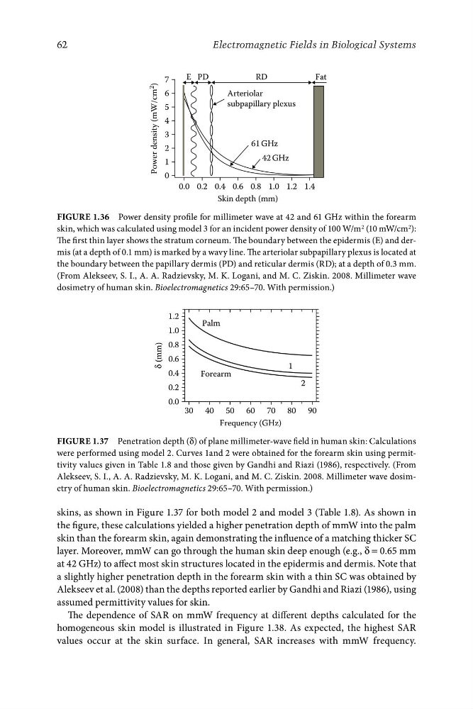
FigurE 1.36 Power density profile for millimeter wave at 42 and 61 GHz within the forearm
skin, which was calculated using model 3 for an incident power density of 100 W/m 2 (10 mW/cm 2 ):
The first thin layer shows the stratum corneum. The boundary between the epidermis (E) and dermis (at a depth of 0.1 mm) is marked by a wavy line. The arteriolar subpapillary plexus is located at
the boundary between the papillary dermis (PD) and reticular dermis (RD); at a depth of 0.3 mm.
(From Alekseev, S. I., A. A. Radzievsky, M. K. Logani, and M. C. Ziskin. 2008. Millimeter wave
dosimetry of human skin. Bioelectromagnetics 29:65–70. With permission.)
1.2
1.0
0.8
0.6
0.4
0.2
δ
(mm)
Palm
Forearm
2
1
0.0
30 40 50 60 70 80 90
Frequency (GHz)
FigurE 1.37 Penetration depth (δ) of plane millimeter-wave field in human skin: Calculations
were performed using model 2. Curves 1and 2 were obtained for the forearm skin using permittivity values given in Table 1.8 and those given by Gandhi and Riazi (1986), respectively. (From
Alekseev, S. I., A. A. Radzievsky, M. K. Logani, and M. C. Ziskin. 2008. Millimeter wave dosimetry of human skin. Bioelectromagnetics 29:65–70. With permission.)
skins, as shown in Figure 1.37 for both model 2 and model 3 (Table 1.8). As shown in
the figure, these calculations yielded a higher penetration depth of mmW into the palm
skin than the forearm skin, again demonstrating the influence of a matching thicker SC
layer. Moreover, mmW can go through the human skin deep enough (e.g., δ = 0.65 mm
at 42 GHz) to affect most skin structures located in the epidermis and dermis. Note that
a slightly higher penetration depth in the forearm skin with a thin SC was obtained by
Alekseev et al. (2008) than the depths reported earlier by Gandhi and Riazi (1986), using
assumed permittivity values for skin.
The dependence of SAR on mmW frequency at different depths calculated for the
homogeneous skin model is illustrated in Figure 1.38. As expected, the highest SAR
values occur at the skin surface. In general, SAR increases with mmW frequency.
RD
Fat
7
Arteriolar
subpapillary plexus
6
5
4
3
2
1
0
61 GHz
42 GHz
Power density (mW/cm
2
)
0.0 0.2 0.4 0.6 0.8 1.0 1.2 1.4
Skin depth (mm)
62
Electromagnetic Fields in Biological Systems
FigurE 1.36 Power density profile for millimeter wave at 42 and 61 GHz within the forearm
skin, which was calculated using model 3 for an incident power density of 100 W/m 2 (10 mW/cm 2 ):
The first thin layer shows the stratum corneum. The boundary between the epidermis (E) and dermis (at a depth of 0.1 mm) is marked by a wavy line. The arteriolar subpapillary plexus is located at
the boundary between the papillary dermis (PD) and reticular dermis (RD); at a depth of 0.3 mm.
(From Alekseev, S. I., A. A. Radzievsky, M. K. Logani, and M. C. Ziskin. 2008. Millimeter wave
dosimetry of human skin. Bioelectromagnetics 29:65–70. With permission.)
1.2
1.0
0.8
0.6
0.4
0.2
δ
(mm)
Palm
Forearm
2
1
0.0
30 40 50 60 70 80 90
Frequency (GHz)
FigurE 1.37 Penetration depth (δ) of plane millimeter-wave field in human skin: Calculations
were performed using model 2. Curves 1and 2 were obtained for the forearm skin using permittivity values given in Table 1.8 and those given by Gandhi and Riazi (1986), respectively. (From
Alekseev, S. I., A. A. Radzievsky, M. K. Logani, and M. C. Ziskin. 2008. Millimeter wave dosimetry of human skin. Bioelectromagnetics 29:65–70. With permission.)
skins, as shown in Figure 1.37 for both model 2 and model 3 (Table 1.8). As shown in
the figure, these calculations yielded a higher penetration depth of mmW into the palm
skin than the forearm skin, again demonstrating the influence of a matching thicker SC
layer. Moreover, mmW can go through the human skin deep enough (e.g., δ = 0.65 mm
at 42 GHz) to affect most skin structures located in the epidermis and dermis. Note that
a slightly higher penetration depth in the forearm skin with a thin SC was obtained by
Alekseev et al. (2008) than the depths reported earlier by Gandhi and Riazi (1986), using
assumed permittivity values for skin.
The dependence of SAR on mmW frequency at different depths calculated for the
homogeneous skin model is illustrated in Figure 1.38. As expected, the highest SAR
values occur at the skin surface. In general, SAR increases with mmW frequency.