

T 1 = 1 microsec
R = − 10 cm
Transmitted pulse (V/m) (×10
3
)
− .20
−.12
−.04
.04
.12
.20
Time (sec) (×10 4 )
R = 10 cm
R = 5 cm
R = − 5 cm
R = 0 cm
−.02
−.01
.02
.01
−.03
.03
51
Coupling of Electromagnetic Fields into Biological Systems
An average conductivity of 0.2 S/m is typical of the conductivities of brain matter in the
frequency range of 10 Hz to 10 MHz (Lin 1976a). The computed results for the constant
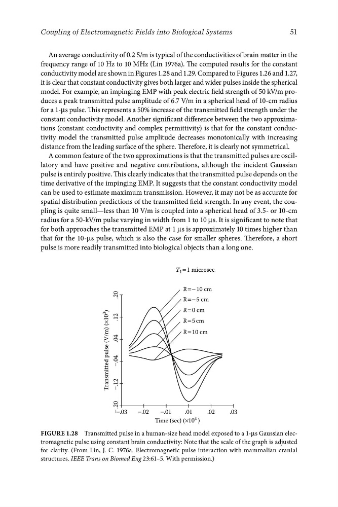
conductivity model are shown in Figures 1.28 and 1.29. Compared to Figures 1.26 and 1.27,
it is clear that constant conductivity gives both larger and wider pulses inside the spherical
model. For example, an impinging EMP with peak electric field strength of 50 kV/m produces a peak transmitted pulse amplitude of 6.7 V/m in a spherical head of 10-cm radius
for a 1-μs pulse. This represents a 50% increase of the transmitted field strength under the
constant conductivity model. Another significant difference between the two approximations (constant conductivity and complex permittivity) is that for the constant conductivity model the transmitted pulse amplitude decreases monotonically with increasing
distance from the leading surface of the sphere. Therefore, it is clearly not symmetrical.
A common feature of the two approximations is that the transmitted pulses are oscillatory and have positive and negative contributions, although the incident Gaussian
pulse is entirely positive. This clearly indicates that the transmitted pulse depends on the
time derivative of the impinging EMP. It suggests that the constant conductivity model
can be used to estimate maximum transmission. However, it may not be as accurate for
spatial distribution predictions of the transmitted field strength. In any event, the coupling is quite small—less than 10 V/m is coupled into a spherical head of 3.5- or 10-cm
radius for a 50-kV/m pulse varying in width from 1 to 10 μs. It is significant to note that
for both approaches the transmitted EMP at 1 μs is approximately 10 times higher than
that for the 10-μs pulse, which is also the case for smaller spheres. Therefore, a short
pulse is more readily transmitted into biological objects than a long one.
FigurE 1.28 Transmitted pulse in a human-size head model exposed to a 1-μs Gaussian electromagnetic pulse using constant brain conductivity: Note that the scale of the graph is adjusted
for clarity. (From Lin, J. C. 1976a. Electromagnetic pulse interaction with mammalian cranial
structures. IEEE Trans on Biomed Eng 23:61–5. With permission.)
R = − 10 cm
Transmitted pulse (V/m) (×10
3
)
− .20
−.12
−.04
.04
.12
.20
Time (sec) (×10 4 )
R = 10 cm
R = 5 cm
R = − 5 cm
R = 0 cm
−.02
−.01
.02
.01
−.03
.03
51
Coupling of Electromagnetic Fields into Biological Systems
An average conductivity of 0.2 S/m is typical of the conductivities of brain matter in the
frequency range of 10 Hz to 10 MHz (Lin 1976a). The computed results for the constant
conductivity model are shown in Figures 1.28 and 1.29. Compared to Figures 1.26 and 1.27,
it is clear that constant conductivity gives both larger and wider pulses inside the spherical
model. For example, an impinging EMP with peak electric field strength of 50 kV/m produces a peak transmitted pulse amplitude of 6.7 V/m in a spherical head of 10-cm radius
for a 1-μs pulse. This represents a 50% increase of the transmitted field strength under the
constant conductivity model. Another significant difference between the two approximations (constant conductivity and complex permittivity) is that for the constant conductivity model the transmitted pulse amplitude decreases monotonically with increasing
distance from the leading surface of the sphere. Therefore, it is clearly not symmetrical.
A common feature of the two approximations is that the transmitted pulses are oscillatory and have positive and negative contributions, although the incident Gaussian
pulse is entirely positive. This clearly indicates that the transmitted pulse depends on the
time derivative of the impinging EMP. It suggests that the constant conductivity model
can be used to estimate maximum transmission. However, it may not be as accurate for
spatial distribution predictions of the transmitted field strength. In any event, the coupling is quite small—less than 10 V/m is coupled into a spherical head of 3.5- or 10-cm
radius for a 50-kV/m pulse varying in width from 1 to 10 μs. It is significant to note that
for both approaches the transmitted EMP at 1 μs is approximately 10 times higher than
that for the 10-μs pulse, which is also the case for smaller spheres. Therefore, a short
pulse is more readily transmitted into biological objects than a long one.
FigurE 1.28 Transmitted pulse in a human-size head model exposed to a 1-μs Gaussian electromagnetic pulse using constant brain conductivity: Note that the scale of the graph is adjusted
for clarity. (From Lin, J. C. 1976a. Electromagnetic pulse interaction with mammalian cranial
structures. IEEE Trans on Biomed Eng 23:61–5. With permission.)