

407
Terahertz Radiation
exposure parameters used in each study: frequency (THz), delivery mode (CW vs. pulsed),
exposure duration, and incubation temperature during exposure. In addition, we provide
a detailed list of the bioeffects that were observed: temperatures generated during exposure, viability, changes in cell growth, and structural/functional changes to lipid membranes, proteins, and DNA. After taking a cursory look at Table 7.2, it is clear that the
exposure parameters, cell type, and end point evaluation techniques used in each study
vary significantly. Therefore, although it may be difficult to draw overarching conclusions
regarding the effects of THz radiation, several effects were consistently observed in many
reports. We shall focus our subsequent discussion on these effects.
7.5.4 Cellular Effects Caused by Terahertz Radiation
In most previous studies, THz radiation did not cause changes at a cellular level (e.g.,
morphology, viability, and growth kinetics) (Scarfi et al. 2003; Zeni et al. 2007; Homenko
et al. 2009; Clothier and Bourne 2003); however, a few studies observed cellular effects.
Specifically, Olshevskaya et al. observed that neurons exhibited morphological changes
when exposed to 2.5 THz radiation (Olshevskaya et al. 2008), and Korenstein-Ilan
et al. observed changes in growth kinetics for cells exposed to THz radiation for long
exposure durations (e.g., 1440 minutes) (Korenstein-Ilan et al. 2008). Our group did
not observe any morphological changes in THz-exposed cells, but we did observe that
the exposed fibroblasts did exhibit enhanced proliferation rates (Wilmink et al. 2010).
A likely explanation for our results is that the exposure conditions we used generated
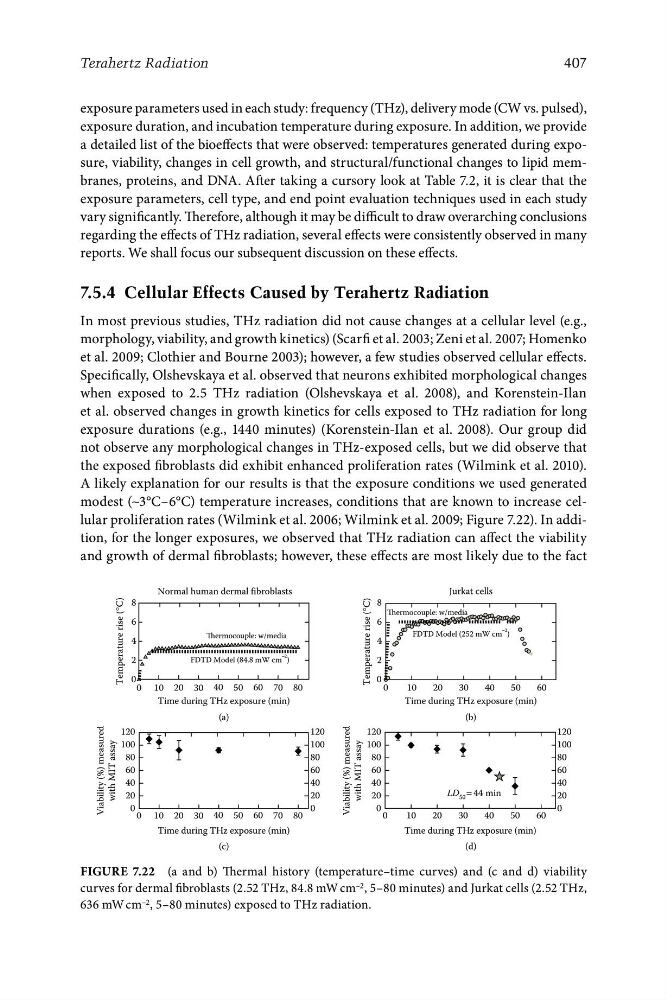
modest (∼3°C–6°C) temperature increases, conditions that are known to increase cellular proliferation rates (Wilmink et al. 2006; Wilmink et al. 2009; Figure 7.22). In addition, for the longer exposures, we observed that THz radiation can affect the viability
and growth of dermal fibroblasts; however, these effects are most likely due to the fact
Normal human dermal fibroblasts
Jurkat cells
Thermocouple: w/media
Thermocouple: w/media
FDTD Model (84.8 mW cm
−2 )
FDTD Model (252 mW cm
−2 )
8
6
4
2
0
8
6
4
2
0
Time during THz exposure (min)
Time during THz exposure (min)
Temperature rise (°C)
Temperature rise (°C)
0 10 20 30 40 50 60
0
10
20
30
40
50
60
0
10
20
LD 50 = 44 min
30
40
50
60
70
Viability (%) measured
with MIT assay
120
100
80
60
40
20
0
120
100
80
60
40
20
0
120
100
80
60
40
20
0
Viability (%) measured
with MIT assay
120
100
80
60
40
20
0
80
(a)
(b)
0 10 20 30 40 50 60 70 80
Time during THz exposure (min)
Time during THz exposure (min)
(c)
(d)
Figure 7.22 (a and b) Thermal history (temperature–time curves) and (c and d) viability
curves for dermal fibroblasts (2.52 THz, 84.8 mW cm –2 , 5–80 minutes) and Jurkat cells (2.52 THz,
636 mW cm –2 , 5–80 minutes) exposed to THz radiation.
Terahertz Radiation
exposure parameters used in each study: frequency (THz), delivery mode (CW vs. pulsed),
exposure duration, and incubation temperature during exposure. In addition, we provide
a detailed list of the bioeffects that were observed: temperatures generated during exposure, viability, changes in cell growth, and structural/functional changes to lipid membranes, proteins, and DNA. After taking a cursory look at Table 7.2, it is clear that the
exposure parameters, cell type, and end point evaluation techniques used in each study
vary significantly. Therefore, although it may be difficult to draw overarching conclusions
regarding the effects of THz radiation, several effects were consistently observed in many
reports. We shall focus our subsequent discussion on these effects.
7.5.4 Cellular Effects Caused by Terahertz Radiation
In most previous studies, THz radiation did not cause changes at a cellular level (e.g.,
morphology, viability, and growth kinetics) (Scarfi et al. 2003; Zeni et al. 2007; Homenko
et al. 2009; Clothier and Bourne 2003); however, a few studies observed cellular effects.
Specifically, Olshevskaya et al. observed that neurons exhibited morphological changes
when exposed to 2.5 THz radiation (Olshevskaya et al. 2008), and Korenstein-Ilan
et al. observed changes in growth kinetics for cells exposed to THz radiation for long
exposure durations (e.g., 1440 minutes) (Korenstein-Ilan et al. 2008). Our group did
not observe any morphological changes in THz-exposed cells, but we did observe that
the exposed fibroblasts did exhibit enhanced proliferation rates (Wilmink et al. 2010).
A likely explanation for our results is that the exposure conditions we used generated
modest (∼3°C–6°C) temperature increases, conditions that are known to increase cellular proliferation rates (Wilmink et al. 2006; Wilmink et al. 2009; Figure 7.22). In addition, for the longer exposures, we observed that THz radiation can affect the viability
and growth of dermal fibroblasts; however, these effects are most likely due to the fact
Normal human dermal fibroblasts
Jurkat cells
Thermocouple: w/media
Thermocouple: w/media
FDTD Model (84.8 mW cm
−2 )
FDTD Model (252 mW cm
−2 )
8
6
4
2
0
8
6
4
2
0
Time during THz exposure (min)
Time during THz exposure (min)
Temperature rise (°C)
Temperature rise (°C)
0 10 20 30 40 50 60
0
10
20
30
40
50
60
0
10
20
LD 50 = 44 min
30
40
50
60
70
Viability (%) measured
with MIT assay
120
100
80
60
40
20
0
120
100
80
60
40
20
0
120
100
80
60
40
20
0
Viability (%) measured
with MIT assay
120
100
80
60
40
20
0
80
(a)
(b)
0 10 20 30 40 50 60 70 80
Time during THz exposure (min)
Time during THz exposure (min)
(c)
(d)
Figure 7.22 (a and b) Thermal history (temperature–time curves) and (c and d) viability
curves for dermal fibroblasts (2.52 THz, 84.8 mW cm –2 , 5–80 minutes) and Jurkat cells (2.52 THz,
636 mW cm –2 , 5–80 minutes) exposed to THz radiation.