

402
Electromagnetic Fields in Biological Systems
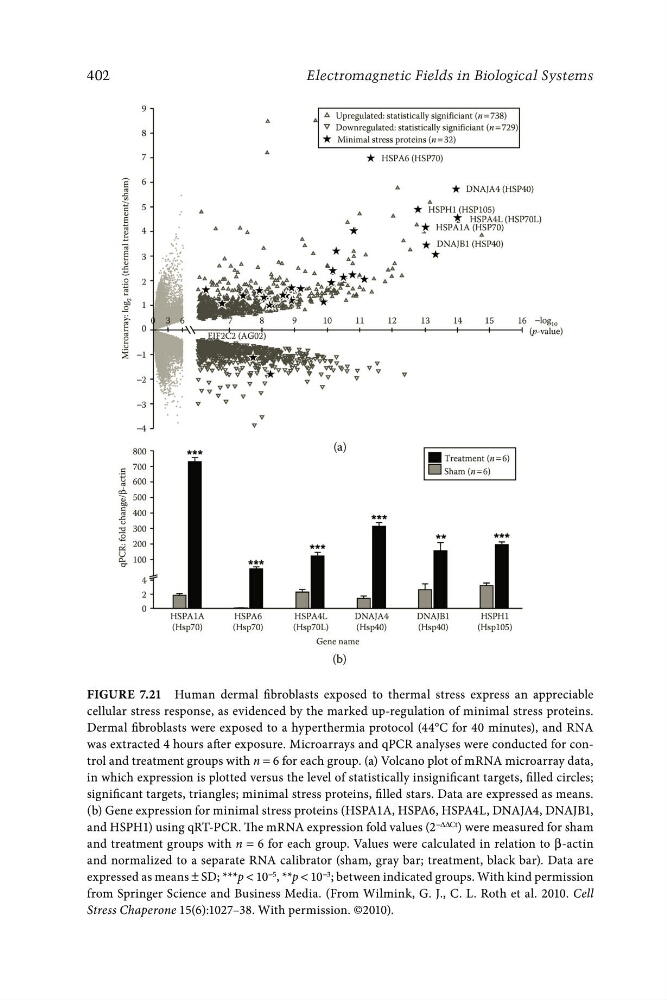
Figure 7.21 Human dermal fibroblasts exposed to thermal stress express an appreciable
cellular stress response, as evidenced by the marked up-regulation of minimal stress proteins.
Dermal fibroblasts were exposed to a hyperthermia protocol (44°C for 40 minutes), and RNA
was extracted 4 hours after exposure. Microarrays and qPCR analyses were conducted for control and treatment groups with n = 6 for each group. (a) Volcano plot of mRNA microarray data,
in which expression is plotted versus the level of statistically insignificant targets, filled circles;
significant targets, triangles; minimal stress proteins, filled stars. Data are expressed as means.
(b) Gene expression for minimal stress proteins (HSPA1A, HSPA6, HSPA4L, DNAJA4, DNAJB1,
and HSPH1) using qRT-PCR. The mRNA expression fold values (2 −ΔΔCt ) were measured for sham
and treatment groups with n = 6 for each group. Values were calculated in relation to β-actin
and normalized to a separate RNA calibrator (sham, gray bar; treatment, black bar). Data are
expressed as means ± SD; ***p < 10 −5 , **p < 10 −3 ; between indicated groups. With kind permission
from Springer Science and Business Media. (From Wilmink, G. J., C. L. Roth et al. 2010. Cell
Stress Chaperone 15(6):1027–38. With permission. ©2010).
Electromagnetic Fields in Biological Systems
Figure 7.21 Human dermal fibroblasts exposed to thermal stress express an appreciable
cellular stress response, as evidenced by the marked up-regulation of minimal stress proteins.
Dermal fibroblasts were exposed to a hyperthermia protocol (44°C for 40 minutes), and RNA
was extracted 4 hours after exposure. Microarrays and qPCR analyses were conducted for control and treatment groups with n = 6 for each group. (a) Volcano plot of mRNA microarray data,
in which expression is plotted versus the level of statistically insignificant targets, filled circles;
significant targets, triangles; minimal stress proteins, filled stars. Data are expressed as means.
(b) Gene expression for minimal stress proteins (HSPA1A, HSPA6, HSPA4L, DNAJA4, DNAJB1,
and HSPH1) using qRT-PCR. The mRNA expression fold values (2 −ΔΔCt ) were measured for sham
and treatment groups with n = 6 for each group. Values were calculated in relation to β-actin
and normalized to a separate RNA calibrator (sham, gray bar; treatment, black bar). Data are
expressed as means ± SD; ***p < 10 −5 , **p < 10 −3 ; between indicated groups. With kind permission
from Springer Science and Business Media. (From Wilmink, G. J., C. L. Roth et al. 2010. Cell
Stress Chaperone 15(6):1027–38. With permission. ©2010).