

(a)
(b)
40 cm
dB scale
0 dB = 0.44e-3 W/kg
−6 −12−18−24
0
5 cm
1 m
3 cm
dB scale
0 dB = 0.12e-3 W/kg
−6 −12 −18 −24
0
−6 −12 −18 −24
0
−6 −12 −18 −24
0
dB scale
0 dB = 0.135 W/kg
20 cm
dB scale
0 dB = 0.54e-3 W/kg
(c)
(d)
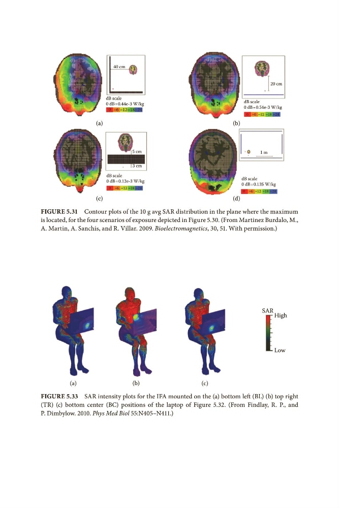
Figure 5.31 Contour plots of the 10 g avg SAR distribution in the plane where the maximum
is located, for the four scenarios of exposure depicted in Figure 5.30. (From Martinez Burdalo, M.,
A. Martin, A. Sanchis, and R. Villar. 2009. Bioelectromagnetics, 30, 51. With permission.)
SAR High
Low
(a)
(b)
(c)
Figure 5.33 SAR intensity plots for the IFA mounted on the (a) bottom left (BL) (b) top right
(TR) (c) bottom center (BC) positions of the laptop of Figure 5.32. (From Findlay, R. P., and
P. Dimbylow. 2010. Phys Med Biol 55:N405–N411.)
(b)
40 cm
dB scale
0 dB = 0.44e-3 W/kg
−6 −12−18−24
0
5 cm
1 m
3 cm
dB scale
0 dB = 0.12e-3 W/kg
−6 −12 −18 −24
0
−6 −12 −18 −24
0
−6 −12 −18 −24
0
dB scale
0 dB = 0.135 W/kg
20 cm
dB scale
0 dB = 0.54e-3 W/kg
(c)
(d)
Figure 5.31 Contour plots of the 10 g avg SAR distribution in the plane where the maximum
is located, for the four scenarios of exposure depicted in Figure 5.30. (From Martinez Burdalo, M.,
A. Martin, A. Sanchis, and R. Villar. 2009. Bioelectromagnetics, 30, 51. With permission.)
SAR High
Low
(a)
(b)
(c)
Figure 5.33 SAR intensity plots for the IFA mounted on the (a) bottom left (BL) (b) top right
(TR) (c) bottom center (BC) positions of the laptop of Figure 5.32. (From Findlay, R. P., and
P. Dimbylow. 2010. Phys Med Biol 55:N405–N411.)