

x
z
y
x
z
y
H
E
x
z
y
Induced electric field lines
17
Coupling of Electromagnetic Fields into Biological Systems
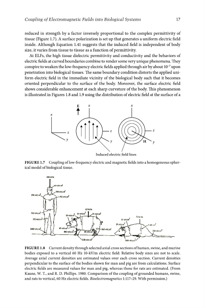
reduced in strength by a factor inversely proportional to the complex permittivity of
tissue (Figure 1.7). A surface polarization is set up that generates a uniform electric field
inside. Although Equation 1.41 suggests that the induced field is independent of body
size, it varies from tissue to tissue as a function of permittivity.
At ELFs, the high tissue dielectric permittivity and conductivity and the behaviors of
electric fields at curved boundaries combine to render some very unique phenomena. They
conspire to weaken the low-frequency electric fields applied through air by about 10 −7 upon
penetration into biological tissues. The same boundary condition distorts the applied uniform electric field in the immediate vicinity of the biological body such that it becomes
oriented perpendicular to the surface of the body. Moreover, the surface electric field
shows considerable enhancement at each sharp curvature of the body. This phenomenon
is illustrated in Figures 1.8 and 1.9 using the distribution of electric field at the surface of a
FigurE 1.7 Coupling of low-frequency electric and magnetic fields into a homogeneous spherical model of biological tissue.
FigurE 1.8 Current density through selected axial cross sections of human, swine, and murine
bodies exposed to a vertical 60 Hz 10-kV/m electric field: Relative body sizes are not to scale.
Average axial current densities are estimated values over each cross section. Current densities
perpendicular to the surface of the bodies shown for man and pig are from calculations. Surface
electric fields are measured values for man and pig, whereas those for rats are estimated. (From
Kaune, W. T., and R. D. Phillips. 1980. Comparison of the coupling of grounded humans, swine,
and rats to vertical, 60 Hz electric fields. Bioelectromagnetics 1:117–29. With permission.)
z
y
x
z
y
H
E
x
z
y
Induced electric field lines
17
Coupling of Electromagnetic Fields into Biological Systems
reduced in strength by a factor inversely proportional to the complex permittivity of
tissue (Figure 1.7). A surface polarization is set up that generates a uniform electric field
inside. Although Equation 1.41 suggests that the induced field is independent of body
size, it varies from tissue to tissue as a function of permittivity.
At ELFs, the high tissue dielectric permittivity and conductivity and the behaviors of
electric fields at curved boundaries combine to render some very unique phenomena. They
conspire to weaken the low-frequency electric fields applied through air by about 10 −7 upon
penetration into biological tissues. The same boundary condition distorts the applied uniform electric field in the immediate vicinity of the biological body such that it becomes
oriented perpendicular to the surface of the body. Moreover, the surface electric field
shows considerable enhancement at each sharp curvature of the body. This phenomenon
is illustrated in Figures 1.8 and 1.9 using the distribution of electric field at the surface of a
FigurE 1.7 Coupling of low-frequency electric and magnetic fields into a homogeneous spherical model of biological tissue.
FigurE 1.8 Current density through selected axial cross sections of human, swine, and murine
bodies exposed to a vertical 60 Hz 10-kV/m electric field: Relative body sizes are not to scale.
Average axial current densities are estimated values over each cross section. Current densities
perpendicular to the surface of the bodies shown for man and pig are from calculations. Surface
electric fields are measured values for man and pig, whereas those for rats are estimated. (From
Kaune, W. T., and R. D. Phillips. 1980. Comparison of the coupling of grounded humans, swine,
and rats to vertical, 60 Hz electric fields. Bioelectromagnetics 1:117–29. With permission.)