

264
Electromagnetic Fields in Biological Systems
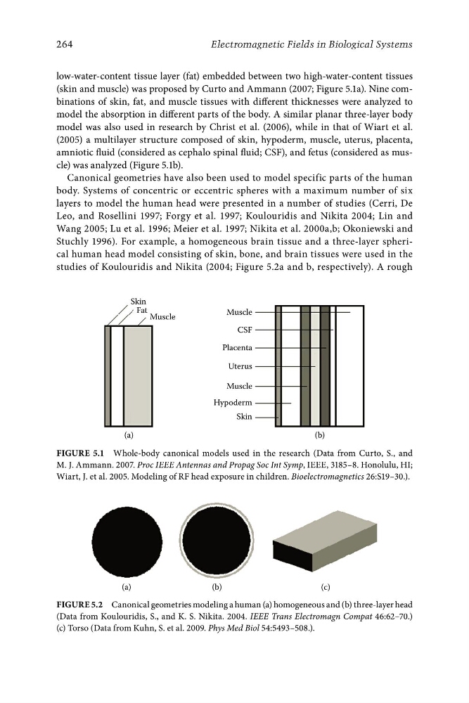
low-water-content tissue layer (fat) embedded between two high-water-content tissues
(skin and muscle) was proposed by Curto and Ammann (2007; Figure 5.1a). Nine combinations of skin, fat, and muscle tissues with different thicknesses were analyzed to
model the absorption in different parts of the body. A similar planar three-layer body
model was also used in research by Christ et al. (2006), while in that of Wiart et al.
(2005) a multilayer structure composed of skin, hypoderm, muscle, uterus, placenta,
amniotic fluid (considered as cephalo spinal fluid; CSF), and fetus (considered as muscle) was analyzed (Figure 5.1b).
Canonical geometries have also been used to model specific parts of the human
body. Systems of concentric or eccentric spheres with a maximum number of six
layers to model the human head were presented in a number of studies (Cerri, De
Leo, and Rosellini 1997; Forgy et al. 1997; Koulouridis and Nikita 2004; Lin and
Wang 2005; Lu et al. 1996; Meier et al. 1997; Nikita et al. 2000a,b; Okoniewski and
Stuchly 1996). For example, a homogeneous brain tissue and a three-layer spherical human head model consisting of skin, bone, and brain tissues were used in the
studies of Koulouridis and Nikita (2004; Figure 5.2a and b, respectively). A rough
Skin
Fat
Muscle
Muscle
CSF
Placenta
Uterus
Muscle
Hypoderm
Skin
(a)
(b)
Figure 5.1 Whole-body canonical models used in the research (Data from Curto, S., and
M. J. Ammann. 2007. Proc IEEE Antennas and Propag Soc Int Symp, IEEE, 3185–8. Honolulu, HI;
Wiart, J. et al. 2005. Modeling of RF head exposure in children. Bioelectromagnetics 26:S19–30.).
(a)
(b)
(c)
Figure 5.2 Canonical geometries modeling a human (a) homogeneous and (b) three-layer head
(Data from Koulouridis, S., and K. S. Nikita. 2004. IEEE Trans Electromagn Compat 46:62–70.)
(c) Torso (Data from Kuhn, S. et al. 2009. Phys Med Biol 54:5493–508.).