

206
Electromagnetic Fields in Biological Systems
E o
2b
+
+
+
+
+
+
+
+
+
2a
J
E 1
E o
E 1
J
I 8
J o
E o
Q o
y
I oc
m
1.7
A
B
C
D
E
F
G
H
0.1
0.09
0.06
0.27
0.12
0.12
0.06
0.05
0.30
0.31
0.30
0.35
0.09
Unit: m
0.22
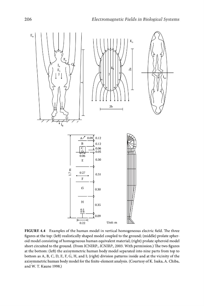
Figure 4.4 Examples of the human model in vertical homogeneous electric field. The three
figures at the top: (left) realistically shaped model coupled to the ground; (middle) prolate spheroid model consisting of homogeneous human equivalent material; (right) prolate spheroid model
short circuited to the ground. (From ICNIRP., ICNIRP., 2003. With permission.) The two figures
at the bottom: (left) the axisymmetric human body model separated into nine parts from top to
bottom as A, B, C, D, E, F, G, H, and I; (right) division patterns inside and at the vicinity of the
axisymmetric human body model for the finite-element analysis. (Courtesy of K. Isaka, A. Chiba,
and W. T. Kaune 1998.)
Electromagnetic Fields in Biological Systems
E o
2b
+
+
+
+
+
+
+
+
+
2a
J
E 1
E o
E 1
J
I 8
J o
E o
Q o
y
I oc
m
1.7
A
B
C
D
E
F
G
H
0.1
0.09
0.06
0.27
0.12
0.12
0.06
0.05
0.30
0.31
0.30
0.35
0.09
Unit: m
0.22
Figure 4.4 Examples of the human model in vertical homogeneous electric field. The three
figures at the top: (left) realistically shaped model coupled to the ground; (middle) prolate spheroid model consisting of homogeneous human equivalent material; (right) prolate spheroid model
short circuited to the ground. (From ICNIRP., ICNIRP., 2003. With permission.) The two figures
at the bottom: (left) the axisymmetric human body model separated into nine parts from top to
bottom as A, B, C, D, E, F, G, H, and I; (right) division patterns inside and at the vicinity of the
axisymmetric human body model for the finite-element analysis. (Courtesy of K. Isaka, A. Chiba,
and W. T. Kaune 1998.)