

97
Pulsed Electric Fields in Biological Cells and Membranes
20
0
0
20
40
60
80
100
40
Time (ns)
Temperature change (K)
θ (degree)
60
80
0
1
2
3
4
5
6
7
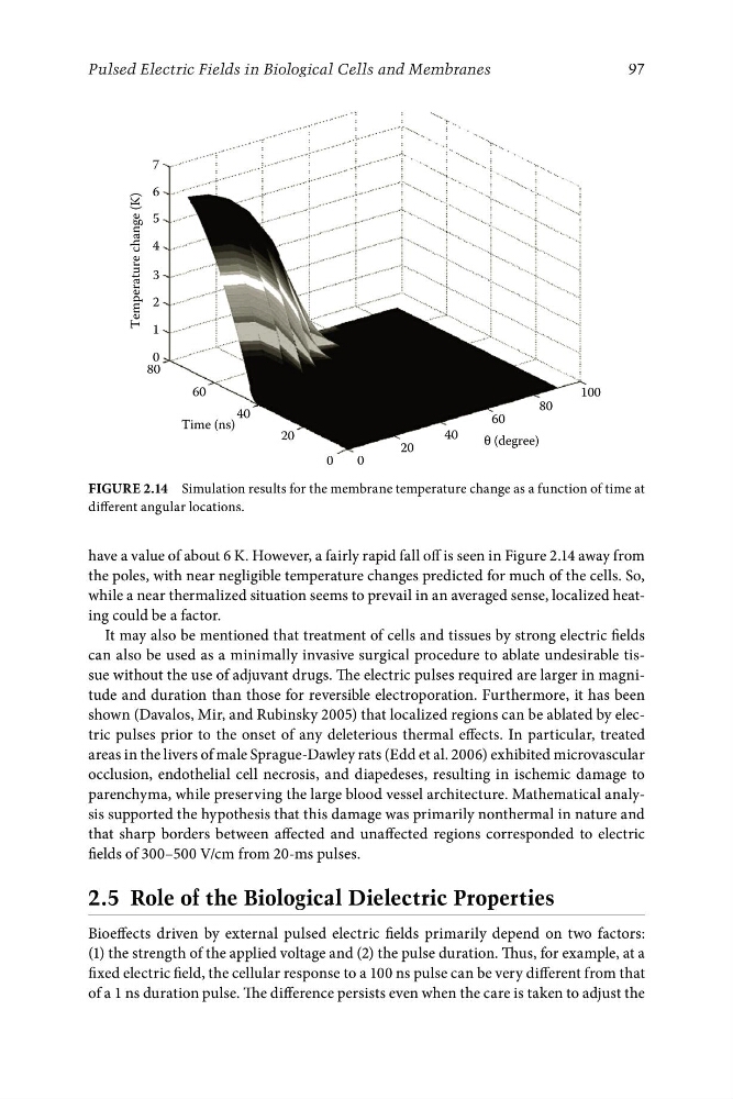
Figure 2.14 Simulation results for the membrane temperature change as a function of time at
different angular locations.
have a value of about 6 K. However, a fairly rapid fall off is seen in Figure 2.14 away from
the poles, with near negligible temperature changes predicted for much of the cells. So,
while a near thermalized situation seems to prevail in an averaged sense, localized heating could be a factor.
It may also be mentioned that treatment of cells and tissues by strong electric fields
can also be used as a minimally invasive surgical procedure to ablate undesirable tissue without the use of adjuvant drugs. The electric pulses required are larger in magnitude and duration than those for reversible electroporation. Furthermore, it has been
shown (Davalos, Mir, and Rubinsky 2005) that localized regions can be ablated by electric pulses prior to the onset of any deleterious thermal effects. In particular, treated
areas in the livers of male Sprague-Dawley rats (Edd et al. 2006) exhibited microvascular
occlusion, endothelial cell necrosis, and diapedeses, resulting in ischemic damage to
parenchyma, while preserving the large blood vessel architecture. Mathematical analysis supported the hypothesis that this damage was primarily nonthermal in nature and
that sharp borders between affected and unaffected regions corresponded to electric
fields of 300–500 V/cm from 20-ms pulses.
2.5 Role of the Biological Dielectric Properties
Bioeffects driven by external pulsed electric fields primarily depend on two factors:
(1) the strength of the applied voltage and (2) the pulse duration. Thus, for example, at a
fixed electric field, the cellular response to a 100 ns pulse can be very different from that
of a 1 ns duration pulse. The difference persists even when the care is taken to adjust the