

236
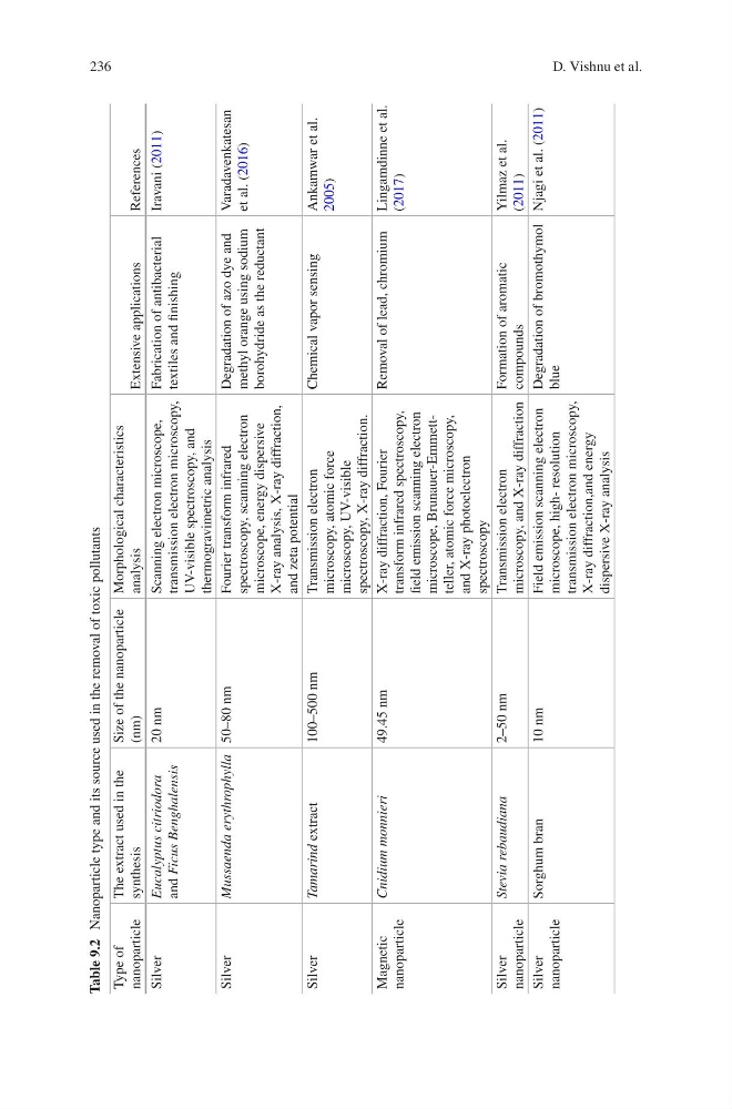
Table 9.2 Nanoparticle type and its source used in the removal of toxic pollutants
Type of
nanoparticle
The extract used in the
synthesis
Size of the nanoparticle
(nm)
Morphological characteristics
analysis
Extensive applications
References
Silver
Eucalyptus citriodora
and Ficus Benghalensis
20 nm
Scanning electron microscope,
transmission electron microscopy,
UV-visible spectroscopy, and
thermogravimetric analysis
Fabrication of antibacterial
textiles and finishing
Iravani (2011)
Silver
Mussaenda erythrophylla
50–80 nm
Fourier transform infrared
spectroscopy, scanning electron
microscope, energy dispersive
X-ray analysis, X-ray diffraction,
and zeta potential
Degradation of azo dye and
methyl orange using sodium
borohydride as the reductant
Varadavenkatesan
et al. (2016)
Silver
Tamarind
extract
100–500 nm
Transmission electron
microscopy, atomic force
microscopy, UV-visible
spectroscopy, X-ray diffraction.
Chemical vapor sensing
Ankamwar et al.
2005)
Magnetic
nanoparticle
Cnidium monnieri
49.45 nm
X-ray diffraction, Fourier
transform infrared spectroscopy,
field emission scanning electron
microscope, Brunauer-Emmettteller, atomic force microscopy,
and X-ray photoelectron
spectroscopy
Removal of lead, chromium Lingamdinne et al.
(2017)
Silver
nanoparticle
Stevia rebaudiana
2–50 nm
Transmission electron
microscopy, and X-ray diffraction
Formation of aromatic
compounds
Yilmaz et al.
(2011)
Silver
nanoparticle
Sorghum bran
10 nm
Field emission scanning electron
microscope, high- resolution
transmission electron microscopy,
X-ray diffraction,and energy
dispersive X-ray analysis
Degradation of bromothymol
blue
Njagi et al. (2011)
D. Vishnu et al.
Table 9.2 Nanoparticle type and its source used in the removal of toxic pollutants
Type of
nanoparticle
The extract used in the
synthesis
Size of the nanoparticle
(nm)
Morphological characteristics
analysis
Extensive applications
References
Silver
Eucalyptus citriodora
and Ficus Benghalensis
20 nm
Scanning electron microscope,
transmission electron microscopy,
UV-visible spectroscopy, and
thermogravimetric analysis
Fabrication of antibacterial
textiles and finishing
Iravani (2011)
Silver
Mussaenda erythrophylla
50–80 nm
Fourier transform infrared
spectroscopy, scanning electron
microscope, energy dispersive
X-ray analysis, X-ray diffraction,
and zeta potential
Degradation of azo dye and
methyl orange using sodium
borohydride as the reductant
Varadavenkatesan
et al. (2016)
Silver
Tamarind
extract
100–500 nm
Transmission electron
microscopy, atomic force
microscopy, UV-visible
spectroscopy, X-ray diffraction.
Chemical vapor sensing
Ankamwar et al.
2005)
Magnetic
nanoparticle
Cnidium monnieri
49.45 nm
X-ray diffraction, Fourier
transform infrared spectroscopy,
field emission scanning electron
microscope, Brunauer-Emmettteller, atomic force microscopy,
and X-ray photoelectron
spectroscopy
Removal of lead, chromium Lingamdinne et al.
(2017)
Silver
nanoparticle
Stevia rebaudiana
2–50 nm
Transmission electron
microscopy, and X-ray diffraction
Formation of aromatic
compounds
Yilmaz et al.
(2011)
Silver
nanoparticle
Sorghum bran
10 nm
Field emission scanning electron
microscope, high- resolution
transmission electron microscopy,
X-ray diffraction,and energy
dispersive X-ray analysis
Degradation of bromothymol
blue
Njagi et al. (2011)
D. Vishnu et al.