

8.3 Self-Reinforcement Behavior of NR
157
Table 8.1 Compounding
recipe of carbon black-loaded
natural rubber for
vulcanization in phr (from
Table 1 in Ref. [88])
Natural rubber (NR)
100
Sulfur
3
Accelerator
4
Zinc oxide
9.85
Stearic acid
3
Carbon black
34
Oil
3
Antioxidant
2
Further, the studies on fatigue behavior of NR continue by using X-ray from
synchrotron sources. For example, the result described in Ref. [88] is introduced
here. The compounding recipe for vulcanization of NR specimen is shown in Table
8.1, which is somewhat similar to the practical formulations including CB, but with
a little increased amount of zinc oxide than usual. The specimen of a cylindrical
shape was used, which had been subjected to the repeated deformations to show
a crack on the surface, before the fatigue test. The crack growth was observed by
scanning electron microscopy (SEM) technique, and the fractographical analysis
was conducted on the results: The section was created by microcutting using SEM
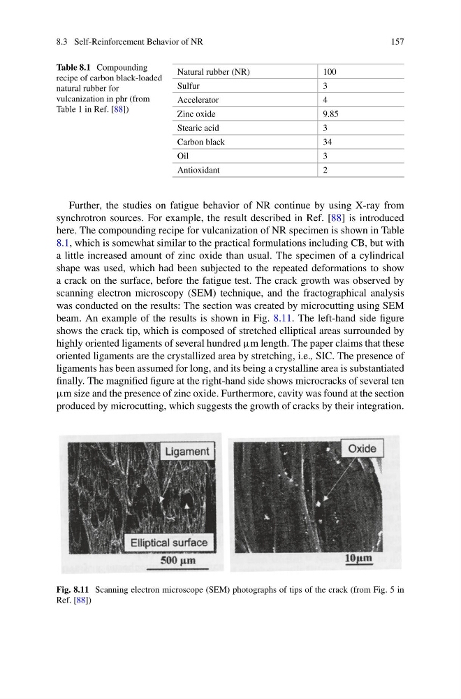
beam. An example of the results is shown in Fig. 8.11. The left-hand side figure
shows the crack tip, which is composed of stretched elliptical areas surrounded by
highly oriented ligaments of several hundred μm length. The paper claims that these
oriented ligaments are the crystallized area by stretching, i.e., SIC. The presence of
ligaments has been assumed for long, and its being a crystalline area is substantiated
finally. The magnified figure at the right-hand side shows microcracks of several ten
μm size and the presence of zinc oxide. Furthermore, cavity was found at the section
produced by microcutting, which suggests the growth of cracks by their integration.
Fig. 8.11 Scanning electron microscope (SEM) photographs of tips of the crack (from Fig. 5 in
Ref. [88])
157
Table 8.1 Compounding
recipe of carbon black-loaded
natural rubber for
vulcanization in phr (from
Table 1 in Ref. [88])
Natural rubber (NR)
100
Sulfur
3
Accelerator
4
Zinc oxide
9.85
Stearic acid
3
Carbon black
34
Oil
3
Antioxidant
2
Further, the studies on fatigue behavior of NR continue by using X-ray from
synchrotron sources. For example, the result described in Ref. [88] is introduced
here. The compounding recipe for vulcanization of NR specimen is shown in Table
8.1, which is somewhat similar to the practical formulations including CB, but with
a little increased amount of zinc oxide than usual. The specimen of a cylindrical
shape was used, which had been subjected to the repeated deformations to show
a crack on the surface, before the fatigue test. The crack growth was observed by
scanning electron microscopy (SEM) technique, and the fractographical analysis
was conducted on the results: The section was created by microcutting using SEM
beam. An example of the results is shown in Fig. 8.11. The left-hand side figure
shows the crack tip, which is composed of stretched elliptical areas surrounded by
highly oriented ligaments of several hundred μm length. The paper claims that these
oriented ligaments are the crystallized area by stretching, i.e., SIC. The presence of
ligaments has been assumed for long, and its being a crystalline area is substantiated
finally. The magnified figure at the right-hand side shows microcracks of several ten
μm size and the presence of zinc oxide. Furthermore, cavity was found at the section
produced by microcutting, which suggests the growth of cracks by their integration.
Fig. 8.11 Scanning electron microscope (SEM) photographs of tips of the crack (from Fig. 5 in
Ref. [88])