

402
region of GoM (2012–2017) at sites located proximate to the DWH wellhead were
analyzed for histopathological changes and disease manifestations (Pulster et al.
2018). Preliminary analysis of hepatic tissues, revealed that 99% exhibited morphological and/or pathological changes, including a spectrum of alterations such as
vacuolar change (lipid and glycogen type), hepatocellular atrophy, lymphocytic
inflammation, migrating larval nematodes, adult nematodes, and intra-biliary myxozoans. In the SGoM Ixtoc 1 spill area, red snapper livers also showed a high incidence (>80%) of a variety of hepatic parenchymal changes. Red snapper liver
variances were found along the whole SGoM, and more than 85% of these pathologies could be related to hydrocarbons among other agents (Fig. 24.8). In contrast,
90% of fish gonad tissues were found to be healthy which might indicate the preservation of reproductive potential (Gracia et al. 2017). In the SGoM, an analysis of
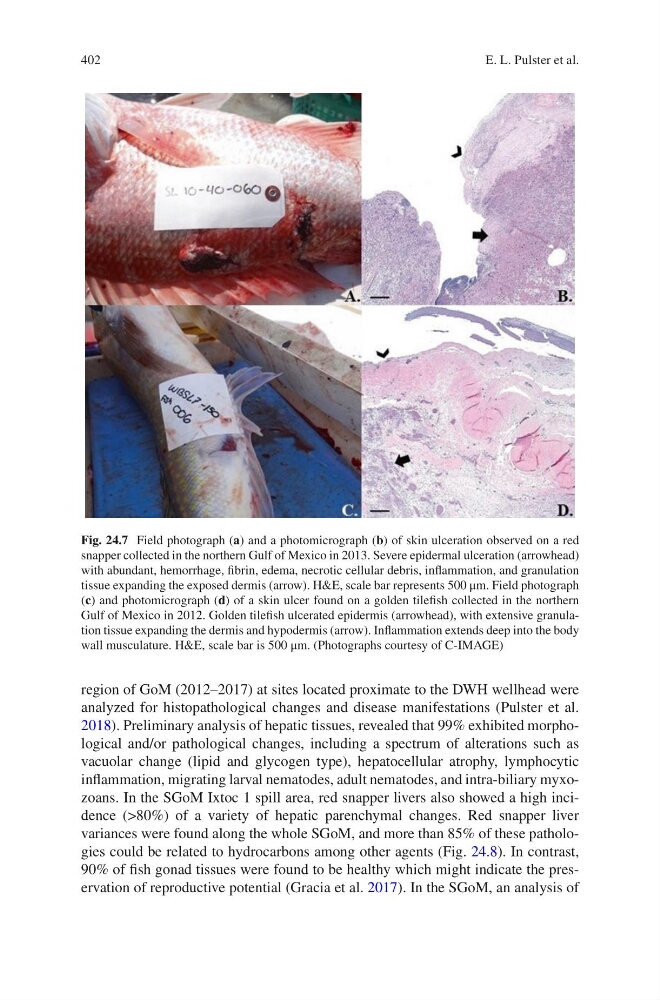
Fig. 24.7 Field photograph (a) and a photomicrograph (b) of skin ulceration observed on a red
snapper collected in the northern Gulf of Mexico in 2013. Severe epidermal ulceration (arrowhead)
with abundant, hemorrhage, fibrin, edema, necrotic cellular debris, inflammation, and granulation
tissue expanding the exposed dermis (arrow). H&E, scale bar represents 500 μm. Field photograph
(c) and photomicrograph (d) of a skin ulcer found on a golden tilefish collected in the northern
Gulf of Mexico in 2012. Golden tilefish ulcerated epidermis (arrowhead), with extensive granulation tissue expanding the dermis and hypodermis (arrow). Inflammation extends deep into the body
wall musculature. H&E, scale bar is 500 μm. (Photographs courtesy of C-IMAGE)
E. L. Pulster et al.
region of GoM (2012–2017) at sites located proximate to the DWH wellhead were
analyzed for histopathological changes and disease manifestations (Pulster et al.
2018). Preliminary analysis of hepatic tissues, revealed that 99% exhibited morphological and/or pathological changes, including a spectrum of alterations such as
vacuolar change (lipid and glycogen type), hepatocellular atrophy, lymphocytic
inflammation, migrating larval nematodes, adult nematodes, and intra-biliary myxozoans. In the SGoM Ixtoc 1 spill area, red snapper livers also showed a high incidence (>80%) of a variety of hepatic parenchymal changes. Red snapper liver
variances were found along the whole SGoM, and more than 85% of these pathologies could be related to hydrocarbons among other agents (Fig. 24.8). In contrast,
90% of fish gonad tissues were found to be healthy which might indicate the preservation of reproductive potential (Gracia et al. 2017). In the SGoM, an analysis of
Fig. 24.7 Field photograph (a) and a photomicrograph (b) of skin ulceration observed on a red
snapper collected in the northern Gulf of Mexico in 2013. Severe epidermal ulceration (arrowhead)
with abundant, hemorrhage, fibrin, edema, necrotic cellular debris, inflammation, and granulation
tissue expanding the exposed dermis (arrow). H&E, scale bar represents 500 μm. Field photograph
(c) and photomicrograph (d) of a skin ulcer found on a golden tilefish collected in the northern
Gulf of Mexico in 2012. Golden tilefish ulcerated epidermis (arrowhead), with extensive granulation tissue expanding the dermis and hypodermis (arrow). Inflammation extends deep into the body
wall musculature. H&E, scale bar is 500 μm. (Photographs courtesy of C-IMAGE)
E. L. Pulster et al.