

488
P. Jain and J. F. Xavier
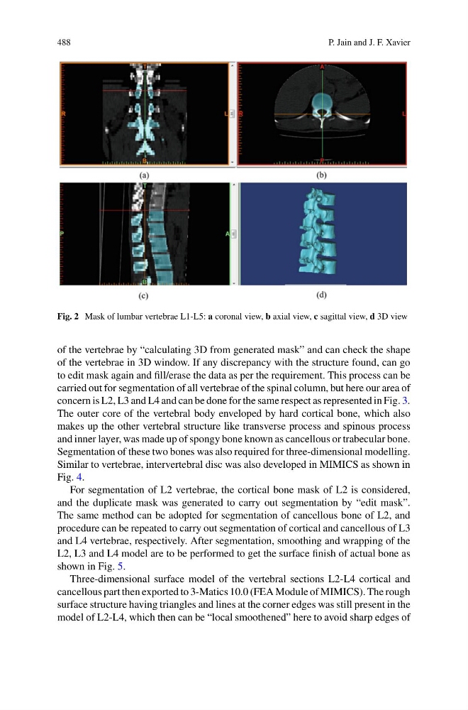
Fig. 2 Mask of lumbar vertebrae L1-L5: a coronal view, b axial view, c sagittal view, d 3D view
of the vertebrae by “calculating 3D from generated mask” and can check the shape
of the vertebrae in 3D window. If any discrepancy with the structure found, can go
to edit mask again and fill/erase the data as per the requirement. This process can be
carried out for segmentation of all vertebrae of the spinal column, but here our area of
concern is L2, L3 and L4 and can be done for the same respect as represented in Fig. 3.
The outer core of the vertebral body enveloped by hard cortical bone, which also
makes up the other vertebral structure like transverse process and spinous process
and inner layer, was made up of spongy bone known as cancellous or trabecular bone.
Segmentation of these two bones was also required for three-dimensional modelling.
Similar to vertebrae, intervertebral disc was also developed in MIMICS as shown in
Fig. 4.
For segmentation of L2 vertebrae, the cortical bone mask of L2 is considered,
and the duplicate mask was generated to carry out segmentation by “edit mask”.
The same method can be adopted for segmentation of cancellous bone of L2, and
procedure can be repeated to carry out segmentation of cortical and cancellous of L3
and L4 vertebrae, respectively. After segmentation, smoothing and wrapping of the
L2, L3 and L4 model are to be performed to get the surface finish of actual bone as
shown in Fig. 5.
Three-dimensional surface model of the vertebral sections L2-L4 cortical and
cancellous part then exported to 3-Matics 10.0 (FEA Module of MIMICS). The rough
surface structure having triangles and lines at the corner edges was still present in the
model of L2-L4, which then can be “local smoothened” here to avoid sharp edges of
P. Jain and J. F. Xavier
Fig. 2 Mask of lumbar vertebrae L1-L5: a coronal view, b axial view, c sagittal view, d 3D view
of the vertebrae by “calculating 3D from generated mask” and can check the shape
of the vertebrae in 3D window. If any discrepancy with the structure found, can go
to edit mask again and fill/erase the data as per the requirement. This process can be
carried out for segmentation of all vertebrae of the spinal column, but here our area of
concern is L2, L3 and L4 and can be done for the same respect as represented in Fig. 3.
The outer core of the vertebral body enveloped by hard cortical bone, which also
makes up the other vertebral structure like transverse process and spinous process
and inner layer, was made up of spongy bone known as cancellous or trabecular bone.
Segmentation of these two bones was also required for three-dimensional modelling.
Similar to vertebrae, intervertebral disc was also developed in MIMICS as shown in
Fig. 4.
For segmentation of L2 vertebrae, the cortical bone mask of L2 is considered,
and the duplicate mask was generated to carry out segmentation by “edit mask”.
The same method can be adopted for segmentation of cancellous bone of L2, and
procedure can be repeated to carry out segmentation of cortical and cancellous of L3
and L4 vertebrae, respectively. After segmentation, smoothing and wrapping of the
L2, L3 and L4 model are to be performed to get the surface finish of actual bone as
shown in Fig. 5.
Three-dimensional surface model of the vertebral sections L2-L4 cortical and
cancellous part then exported to 3-Matics 10.0 (FEA Module of MIMICS). The rough
surface structure having triangles and lines at the corner edges was still present in the
model of L2-L4, which then can be “local smoothened” here to avoid sharp edges of