

A Simplified Method for Conversion …
487
2.2 CT Image Segmentation in Mimics
CT images of the subject were imported in the Materialise’s Interactive Medical
Image Control System (MIMICS) software (17.0, Materialise NV, Belgium) and
used to generate the 3D surface model of the vertebral column L2-L4. Conversion
of CT images into 3D model involves the initial process called “segmentation”.
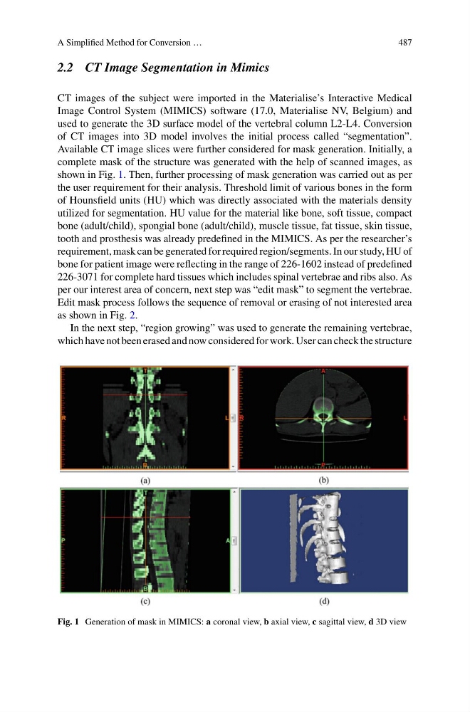
Available CT image slices were further considered for mask generation. Initially, a
complete mask of the structure was generated with the help of scanned images, as
shown in Fig. 1. Then, further processing of mask generation was carried out as per
the user requirement for their analysis. Threshold limit of various bones in the form
of Hounsfield units (HU) which was directly associated with the materials density
utilized for segmentation. HU value for the material like bone, soft tissue, compact
bone (adult/child), spongial bone (adult/child), muscle tissue, fat tissue, skin tissue,
tooth and prosthesis was already predefined in the MIMICS. As per the researcher’s
requirement, mask can be generated for required region/segments. In our study, HU of
bone for patient image were reflecting in the range of 226-1602 instead of predefined
226-3071 for complete hard tissues which includes spinal vertebrae and ribs also. As
per our interest area of concern, next step was “edit mask” to segment the vertebrae.
Edit mask process follows the sequence of removal or erasing of not interested area
as shown in Fig. 2.
In the next step, “region growing” was used to generate the remaining vertebrae,
which have not been erased and now considered for work. User can check the structure
Fig. 1 Generation of mask in MIMICS: a coronal view, b axial view, c sagittal view, d 3D view
487
2.2 CT Image Segmentation in Mimics
CT images of the subject were imported in the Materialise’s Interactive Medical
Image Control System (MIMICS) software (17.0, Materialise NV, Belgium) and
used to generate the 3D surface model of the vertebral column L2-L4. Conversion
of CT images into 3D model involves the initial process called “segmentation”.
Available CT image slices were further considered for mask generation. Initially, a
complete mask of the structure was generated with the help of scanned images, as
shown in Fig. 1. Then, further processing of mask generation was carried out as per
the user requirement for their analysis. Threshold limit of various bones in the form
of Hounsfield units (HU) which was directly associated with the materials density
utilized for segmentation. HU value for the material like bone, soft tissue, compact
bone (adult/child), spongial bone (adult/child), muscle tissue, fat tissue, skin tissue,
tooth and prosthesis was already predefined in the MIMICS. As per the researcher’s
requirement, mask can be generated for required region/segments. In our study, HU of
bone for patient image were reflecting in the range of 226-1602 instead of predefined
226-3071 for complete hard tissues which includes spinal vertebrae and ribs also. As
per our interest area of concern, next step was “edit mask” to segment the vertebrae.
Edit mask process follows the sequence of removal or erasing of not interested area
as shown in Fig. 2.
In the next step, “region growing” was used to generate the remaining vertebrae,
which have not been erased and now considered for work. User can check the structure
Fig. 1 Generation of mask in MIMICS: a coronal view, b axial view, c sagittal view, d 3D view