

8.6 Neural Development
461
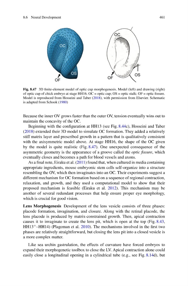
Fig. 8.47 3D finite-element model of optic cup morphogenesis. Model (left) and drawing (right)
of optic cup of chick embryo at stage HH16. OC = optic cup; OS = optic stalk; OF = optic fissure.
Model is reproduced from Hosseini and Taber (2018), with permission from Elsevier. Schematic
is adapted from Schook (1980)
Because the inner OV grows faster than the outer OV, tension eventually wins out to
maintain the concavity of the OC.
Beginning with the configuration at HH13 (see Fig. 8.44c), Hosseini and Taber
(2018) extended their 3D model to simulate OC formation. They added a relatively
stiff matrix layer and prescribed growth in a pattern that is qualitatively consistent
with the axisymmetric model above. At stage HH16, the shape of the OC given
by the model is quite realistic (Fig. 8.47). One unexpected consequence of the
asymmetric geometry is the appearance of a groove called the optic fissure, which
eventually closes and becomes a path for blood vessels and axons.
As a final note, Eiraku et al. (2011) found that, when cultured in media containing
appropriate ingredients, mouse embryonic stem cells self-organize into a structure
resembling the OV, which then invaginates into an OC. Their experiments suggest a
different mechanism for OC formation based on a sequence of regional contraction,
relaxation, and growth, and they used a computational model to show that their
proposed mechanism is feasible (Eiraku et al. 2012). This mechanism may be
another of several redundant processes that help ensure proper eye morphology,
which is crucial for good vision.
Lens Morphogenesis Development of the lens vesicle consists of three phases:
placode formation, invagination, and closure. Along with the retinal placode, the
lens placode is produced by matrix-constrained growth. Then, apical contraction
causes it to invaginate to create the lens pit, which is open at the top (Fig. 8.43,
HH13 + –HH14) (Plageman et al. 2010). The mechanisms involved in the first two
phases are relatively straightforward, but closing the lens pit into a closed vesicle is
a more complex matter.
Like sea urchin gastrulation, the effects of curvature have forced embryos to
expand their morphogenetic toolbox to close the LV. Apical contraction alone could
easily close a longitudinal opening in a cylindrical tube (e.g., see Fig. 8.14d), but
461
Fig. 8.47 3D finite-element model of optic cup morphogenesis. Model (left) and drawing (right)
of optic cup of chick embryo at stage HH16. OC = optic cup; OS = optic stalk; OF = optic fissure.
Model is reproduced from Hosseini and Taber (2018), with permission from Elsevier. Schematic
is adapted from Schook (1980)
Because the inner OV grows faster than the outer OV, tension eventually wins out to
maintain the concavity of the OC.
Beginning with the configuration at HH13 (see Fig. 8.44c), Hosseini and Taber
(2018) extended their 3D model to simulate OC formation. They added a relatively
stiff matrix layer and prescribed growth in a pattern that is qualitatively consistent
with the axisymmetric model above. At stage HH16, the shape of the OC given
by the model is quite realistic (Fig. 8.47). One unexpected consequence of the
asymmetric geometry is the appearance of a groove called the optic fissure, which
eventually closes and becomes a path for blood vessels and axons.
As a final note, Eiraku et al. (2011) found that, when cultured in media containing
appropriate ingredients, mouse embryonic stem cells self-organize into a structure
resembling the OV, which then invaginates into an OC. Their experiments suggest a
different mechanism for OC formation based on a sequence of regional contraction,
relaxation, and growth, and they used a computational model to show that their
proposed mechanism is feasible (Eiraku et al. 2012). This mechanism may be
another of several redundant processes that help ensure proper eye morphology,
which is crucial for good vision.
Lens Morphogenesis Development of the lens vesicle consists of three phases:
placode formation, invagination, and closure. Along with the retinal placode, the
lens placode is produced by matrix-constrained growth. Then, apical contraction
causes it to invaginate to create the lens pit, which is open at the top (Fig. 8.43,
HH13 + –HH14) (Plageman et al. 2010). The mechanisms involved in the first two
phases are relatively straightforward, but closing the lens pit into a closed vesicle is
a more complex matter.
Like sea urchin gastrulation, the effects of curvature have forced embryos to
expand their morphogenetic toolbox to close the LV. Apical contraction alone could
easily close a longitudinal opening in a cylindrical tube (e.g., see Fig. 8.14d), but