

8.5 Early Heart Development
441
Table 8.1 Hamburger–Hamilton stages for chick embryo, approximate incubation times, and
corresponding days of human gestation
Chicken HH stage
6
10
12
16
18
21
24
26
34
36
46
Chicken (days)
1
1.5
2
2.25
3
3.5
4.5
5
8
10
21
Human (days)
20
22
24
28
33
38
41
43
54
58
280
HF
NP
Henson's
node
primitive
streak
anterior
posterior
HF
foregut
vitelline membrane
anterior
posterior
ventral
dorsal
a
a '
a
a'
(a)
(b)
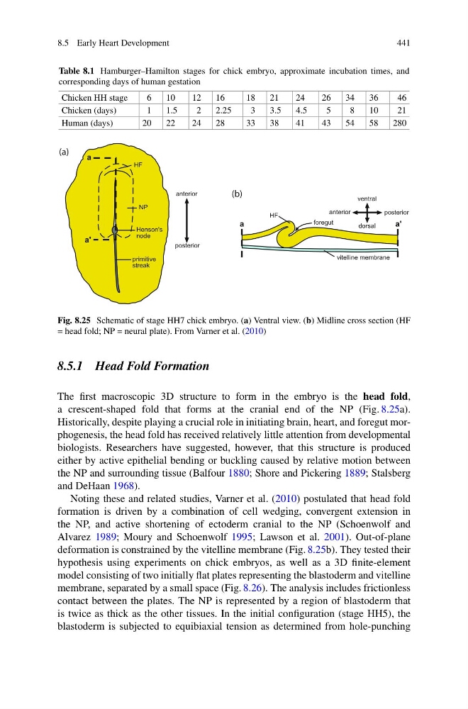
Fig. 8.25 Schematic of stage HH7 chick embryo. (a) Ventral view. (b) Midline cross section (HF
= head fold; NP = neural plate). From Varner et al. (2010)
8.5.1 Head Fold Formation
The first macroscopic 3D structure to form in the embryo is the head fold,
a crescent-shaped fold that forms at the cranial end of the NP (Fig. 8.25a).
Historically, despite playing a crucial role in initiating brain, heart, and foregut morphogenesis, the head fold has received relatively little attention from developmental
biologists. Researchers have suggested, however, that this structure is produced
either by active epithelial bending or buckling caused by relative motion between
the NP and surrounding tissue (Balfour 1880; Shore and Pickering 1889; Stalsberg
and DeHaan 1968).
Noting these and related studies, Varner et al. (2010) postulated that head fold
formation is driven by a combination of cell wedging, convergent extension in
the NP, and active shortening of ectoderm cranial to the NP (Schoenwolf and
Alvarez 1989; Moury and Schoenwolf 1995; Lawson et al. 2001). Out-of-plane
deformation is constrained by the vitelline membrane (Fig. 8.25b). They tested their
hypothesis using experiments on chick embryos, as well as a 3D finite-element
model consisting of two initially flat plates representing the blastoderm and vitelline
membrane, separated by a small space (Fig. 8.26). The analysis includes frictionless
contact between the plates. The NP is represented by a region of blastoderm that
is twice as thick as the other tissues. In the initial configuration (stage HH5), the
blastoderm is subjected to equibiaxial tension as determined from hole-punching
441
Table 8.1 Hamburger–Hamilton stages for chick embryo, approximate incubation times, and
corresponding days of human gestation
Chicken HH stage
6
10
12
16
18
21
24
26
34
36
46
Chicken (days)
1
1.5
2
2.25
3
3.5
4.5
5
8
10
21
Human (days)
20
22
24
28
33
38
41
43
54
58
280
HF
NP
Henson's
node
primitive
streak
anterior
posterior
HF
foregut
vitelline membrane
anterior
posterior
ventral
dorsal
a
a '
a
a'
(a)
(b)
Fig. 8.25 Schematic of stage HH7 chick embryo. (a) Ventral view. (b) Midline cross section (HF
= head fold; NP = neural plate). From Varner et al. (2010)
8.5.1 Head Fold Formation
The first macroscopic 3D structure to form in the embryo is the head fold,
a crescent-shaped fold that forms at the cranial end of the NP (Fig. 8.25a).
Historically, despite playing a crucial role in initiating brain, heart, and foregut morphogenesis, the head fold has received relatively little attention from developmental
biologists. Researchers have suggested, however, that this structure is produced
either by active epithelial bending or buckling caused by relative motion between
the NP and surrounding tissue (Balfour 1880; Shore and Pickering 1889; Stalsberg
and DeHaan 1968).
Noting these and related studies, Varner et al. (2010) postulated that head fold
formation is driven by a combination of cell wedging, convergent extension in
the NP, and active shortening of ectoderm cranial to the NP (Schoenwolf and
Alvarez 1989; Moury and Schoenwolf 1995; Lawson et al. 2001). Out-of-plane
deformation is constrained by the vitelline membrane (Fig. 8.25b). They tested their
hypothesis using experiments on chick embryos, as well as a 3D finite-element
model consisting of two initially flat plates representing the blastoderm and vitelline
membrane, separated by a small space (Fig. 8.26). The analysis includes frictionless
contact between the plates. The NP is represented by a region of blastoderm that
is twice as thick as the other tissues. In the initial configuration (stage HH5), the
blastoderm is subjected to equibiaxial tension as determined from hole-punching