

438
8 Morphogenesis
layer (Fig. 8.22d). Experiments on sea urchin embryos suggest that the apical
lamina can swell, causing the epithelium to bend inward, as in Example 8.3 (Lane
1993). According to the computational model of Davidson et al. (1995), realistic
invagination requires the hyaline layer to be stiffer than the apical lamina, and
both matrix layers should be stiffer than the cells. Experimental measurements are
consistent with this finding (Davidson et al. 1999).
As a first approximation, the apical lamina (c = 50x endoderm modulus) and
hyaline layer (c = 200x endoderm modulus) are represented by two relatively thin
outer layers of the inner region (Fig. 8.22d). Isotropic swelling of the apical lamina
is simulated by specifying G r = G > 1. Consistent with the model of Davidson
et al. (1995), results for G = 1.6 show that the invagination is wider and flatter,
and the edge sharper than those generated by apical constriction (Fig. 8.22e, f). This
morphology agrees reasonably well with gastrulation in some types of sea urchin
(Kominami and Takata 2004). Overall, published studies of sea urchin gastrulation
seem to support matrix swelling over apical constriction (Davidson et al. 1999).
8.4.4 Gastrulation in Sea Squirts
Sea squirts (ascidians) are animals found attached to solid surfaces under water
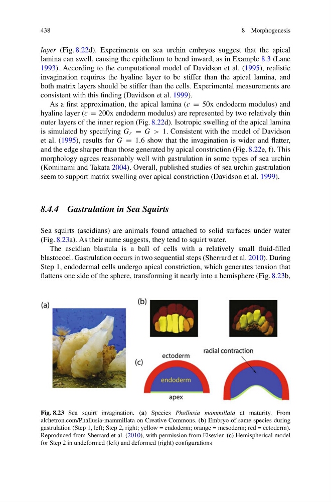
(Fig. 8.23a). As their name suggests, they tend to squirt water.
The ascidian blastula is a ball of cells with a relatively small fluid-filled
blastocoel. Gastrulation occurs in two sequential steps (Sherrard et al. 2010). During
Step 1, endodermal cells undergo apical constriction, which generates tension that
flattens one side of the sphere, transforming it nearly into a hemisphere (Fig. 8.23b,
Fig. 8.23 Sea squirt invagination. (a) Species Phallusia mammillata at maturity. From
alchetron.com/Phallusia-mammillata on Creative Commons. (b) Embryo of same species during
gastrulation (Step 1, left; Step 2, right; yellow = endoderm; orange = mesoderm; red = ectoderm).
Reproduced from Sherrard et al. (2010), with permission from Elsevier. (c) Hemispherical model
for Step 2 in undeformed (left) and deformed (right) configurations
8 Morphogenesis
layer (Fig. 8.22d). Experiments on sea urchin embryos suggest that the apical
lamina can swell, causing the epithelium to bend inward, as in Example 8.3 (Lane
1993). According to the computational model of Davidson et al. (1995), realistic
invagination requires the hyaline layer to be stiffer than the apical lamina, and
both matrix layers should be stiffer than the cells. Experimental measurements are
consistent with this finding (Davidson et al. 1999).
As a first approximation, the apical lamina (c = 50x endoderm modulus) and
hyaline layer (c = 200x endoderm modulus) are represented by two relatively thin
outer layers of the inner region (Fig. 8.22d). Isotropic swelling of the apical lamina
is simulated by specifying G r = G > 1. Consistent with the model of Davidson
et al. (1995), results for G = 1.6 show that the invagination is wider and flatter,
and the edge sharper than those generated by apical constriction (Fig. 8.22e, f). This
morphology agrees reasonably well with gastrulation in some types of sea urchin
(Kominami and Takata 2004). Overall, published studies of sea urchin gastrulation
seem to support matrix swelling over apical constriction (Davidson et al. 1999).
8.4.4 Gastrulation in Sea Squirts
Sea squirts (ascidians) are animals found attached to solid surfaces under water
(Fig. 8.23a). As their name suggests, they tend to squirt water.
The ascidian blastula is a ball of cells with a relatively small fluid-filled
blastocoel. Gastrulation occurs in two sequential steps (Sherrard et al. 2010). During
Step 1, endodermal cells undergo apical constriction, which generates tension that
flattens one side of the sphere, transforming it nearly into a hemisphere (Fig. 8.23b,
Fig. 8.23 Sea squirt invagination. (a) Species Phallusia mammillata at maturity. From
alchetron.com/Phallusia-mammillata on Creative Commons. (b) Embryo of same species during
gastrulation (Step 1, left; Step 2, right; yellow = endoderm; orange = mesoderm; red = ectoderm).
Reproduced from Sherrard et al. (2010), with permission from Elsevier. (c) Hemispherical model
for Step 2 in undeformed (left) and deformed (right) configurations