

7.6 Case Study: Growth and Remodeling of Arteries
371
remodeling of a mature artery during health and disease. Two problems are
considered. The first deals with the effects of matrix remodeling on residual stress
and opening angles. The second problem involves the development of an aneurysm.
7.6.1 Residual Stress and Opening Angles
As discussed in Sects. 6.7 and 6.11.5, the mechanics that determine the magnitude
and evolution of opening angles in arteries can be quite complex. This is especially
true when multiple constituents are involved. The main objective here is to gain
insight into how the extracellular matrix affects residual stress and opening angles.
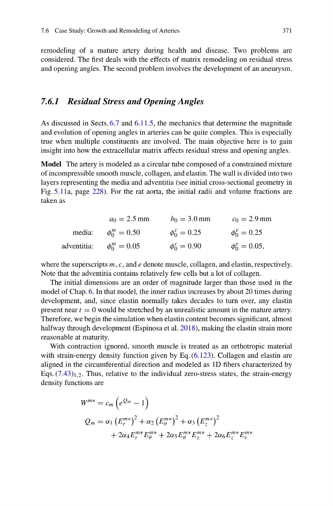
Model The artery is modeled as a circular tube composed of a constrained mixture
of incompressible smooth muscle, collagen, and elastin. The wall is divided into two
layers representing the media and adventitia (see initial cross-sectional geometry in
Fig. 5.11a, page 228). For the rat aorta, the initial radii and volume fractions are
taken as
a 0 = 2.5 mm
b 0 = 3.0 mm
c 0 = 2.9 mm
media:
φ
m
0 = 0.50
φ
c
0 = 0.25
φ
e
0 = 0.25
adventitia:
φ
m
0 = 0.05
φ
c
0 = 0.90
φ
e
0 = 0.05,
where the superscripts m, c, and e denote muscle, collagen, and elastin, respectively.
Note that the adventitia contains relatively few cells but a lot of collagen.
The initial dimensions are an order of magnitude larger than those used in the
model of Chap. 6. In that model, the inner radius increases by about 20 times during
development, and, since elastin normally takes decades to turn over, any elastin
present near t = 0 would be stretched by an unrealistic amount in the mature artery.
Therefore, we begin the simulation when elastin content becomes significant, almost
halfway through development (Espinosa et al. 2018), making the elastin strain more
reasonable at maturity.
With contraction ignored, smooth muscle is treated as an orthotropic material
with strain-energy density function given by Eq. (6.123). Collagen and elastin are
aligned in the circumferential direction and modeled as 1D fibers characterized by
Eqs. (7.43) 1,2 . Thus, relative to the individual zero-stress states, the strain-energy
density functions are
W
m∗
= c m
e
Q m − 1
Q m = α 1
E
m∗
r
2 + α 2
E
m∗
θ
2 + α 3
E
m∗
z
2
+ 2α 4 E
m∗
r E
m∗
θ + 2α 5 E
m∗
θ E
m∗
z + 2α 6 E
m∗
z E
m∗
r
371
remodeling of a mature artery during health and disease. Two problems are
considered. The first deals with the effects of matrix remodeling on residual stress
and opening angles. The second problem involves the development of an aneurysm.
7.6.1 Residual Stress and Opening Angles
As discussed in Sects. 6.7 and 6.11.5, the mechanics that determine the magnitude
and evolution of opening angles in arteries can be quite complex. This is especially
true when multiple constituents are involved. The main objective here is to gain
insight into how the extracellular matrix affects residual stress and opening angles.
Model The artery is modeled as a circular tube composed of a constrained mixture
of incompressible smooth muscle, collagen, and elastin. The wall is divided into two
layers representing the media and adventitia (see initial cross-sectional geometry in
Fig. 5.11a, page 228). For the rat aorta, the initial radii and volume fractions are
taken as
a 0 = 2.5 mm
b 0 = 3.0 mm
c 0 = 2.9 mm
media:
φ
m
0 = 0.50
φ
c
0 = 0.25
φ
e
0 = 0.25
adventitia:
φ
m
0 = 0.05
φ
c
0 = 0.90
φ
e
0 = 0.05,
where the superscripts m, c, and e denote muscle, collagen, and elastin, respectively.
Note that the adventitia contains relatively few cells but a lot of collagen.
The initial dimensions are an order of magnitude larger than those used in the
model of Chap. 6. In that model, the inner radius increases by about 20 times during
development, and, since elastin normally takes decades to turn over, any elastin
present near t = 0 would be stretched by an unrealistic amount in the mature artery.
Therefore, we begin the simulation when elastin content becomes significant, almost
halfway through development (Espinosa et al. 2018), making the elastin strain more
reasonable at maturity.
With contraction ignored, smooth muscle is treated as an orthotropic material
with strain-energy density function given by Eq. (6.123). Collagen and elastin are
aligned in the circumferential direction and modeled as 1D fibers characterized by
Eqs. (7.43) 1,2 . Thus, relative to the individual zero-stress states, the strain-energy
density functions are
W
m∗
= c m
e
Q m − 1
Q m = α 1
E
m∗
r
2 + α 2
E
m∗
θ
2 + α 3
E
m∗
z
2
+ 2α 4 E
m∗
r E
m∗
θ + 2α 5 E
m∗
θ E
m∗
z + 2α 6 E
m∗
z E
m∗
r