

7.1 Determinants of Material Properties in Soft Tissues
343
)
b
(
)
a
(
σ
E
elastin
collagen
both
σ
E
increasing time
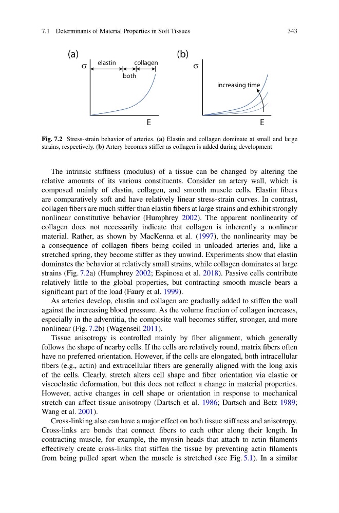
Fig. 7.2 Stress-strain behavior of arteries. (a) Elastin and collagen dominate at small and large
strains, respectively. (b) Artery becomes stiffer as collagen is added during development
The intrinsic stiffness (modulus) of a tissue can be changed by altering the
relative amounts of its various constituents. Consider an artery wall, which is
composed mainly of elastin, collagen, and smooth muscle cells. Elastin fibers
are comparatively soft and have relatively linear stress-strain curves. In contrast,
collagen fibers are much stiffer than elastin fibers at large strains and exhibit strongly
nonlinear constitutive behavior (Humphrey 2002). The apparent nonlinearity of
collagen does not necessarily indicate that collagen is inherently a nonlinear
material. Rather, as shown by MacKenna et al. (1997), the nonlinearity may be
a consequence of collagen fibers being coiled in unloaded arteries and, like a
stretched spring, they become stiffer as they unwind. Experiments show that elastin
dominates the behavior at relatively small strains, while collagen dominates at large
strains (Fig. 7.2a) (Humphrey 2002; Espinosa et al. 2018). Passive cells contribute
relatively little to the global properties, but contracting smooth muscle bears a
significant part of the load (Faury et al. 1999).
As arteries develop, elastin and collagen are gradually added to stiffen the wall
against the increasing blood pressure. As the volume fraction of collagen increases,
especially in the adventitia, the composite wall becomes stiffer, stronger, and more
nonlinear (Fig. 7.2b) (Wagenseil 2011).
Tissue anisotropy is controlled mainly by fiber alignment, which generally
follows the shape of nearby cells. If the cells are relatively round, matrix fibers often
have no preferred orientation. However, if the cells are elongated, both intracellular
fibers (e.g., actin) and extracellular fibers are generally aligned with the long axis
of the cells. Clearly, stretch alters cell shape and fiber orientation via elastic or
viscoelastic deformation, but this does not reflect a change in material properties.
However, active changes in cell shape or orientation in response to mechanical
stretch can affect tissue anisotropy (Dartsch et al. 1986; Dartsch and Betz 1989;
Wang et al. 2001).
Cross-linking also can have a major effect on both tissue stiffness and anisotropy.
Cross-links are bonds that connect fibers to each other along their length. In
contracting muscle, for example, the myosin heads that attach to actin filaments
effectively create cross-links that stiffen the tissue by preventing actin filaments
from being pulled apart when the muscle is stretched (see Fig. 5.1). In a similar
343
)
b
(
)
a
(
σ
E
elastin
collagen
both
σ
E
increasing time
Fig. 7.2 Stress-strain behavior of arteries. (a) Elastin and collagen dominate at small and large
strains, respectively. (b) Artery becomes stiffer as collagen is added during development
The intrinsic stiffness (modulus) of a tissue can be changed by altering the
relative amounts of its various constituents. Consider an artery wall, which is
composed mainly of elastin, collagen, and smooth muscle cells. Elastin fibers
are comparatively soft and have relatively linear stress-strain curves. In contrast,
collagen fibers are much stiffer than elastin fibers at large strains and exhibit strongly
nonlinear constitutive behavior (Humphrey 2002). The apparent nonlinearity of
collagen does not necessarily indicate that collagen is inherently a nonlinear
material. Rather, as shown by MacKenna et al. (1997), the nonlinearity may be
a consequence of collagen fibers being coiled in unloaded arteries and, like a
stretched spring, they become stiffer as they unwind. Experiments show that elastin
dominates the behavior at relatively small strains, while collagen dominates at large
strains (Fig. 7.2a) (Humphrey 2002; Espinosa et al. 2018). Passive cells contribute
relatively little to the global properties, but contracting smooth muscle bears a
significant part of the load (Faury et al. 1999).
As arteries develop, elastin and collagen are gradually added to stiffen the wall
against the increasing blood pressure. As the volume fraction of collagen increases,
especially in the adventitia, the composite wall becomes stiffer, stronger, and more
nonlinear (Fig. 7.2b) (Wagenseil 2011).
Tissue anisotropy is controlled mainly by fiber alignment, which generally
follows the shape of nearby cells. If the cells are relatively round, matrix fibers often
have no preferred orientation. However, if the cells are elongated, both intracellular
fibers (e.g., actin) and extracellular fibers are generally aligned with the long axis
of the cells. Clearly, stretch alters cell shape and fiber orientation via elastic or
viscoelastic deformation, but this does not reflect a change in material properties.
However, active changes in cell shape or orientation in response to mechanical
stretch can affect tissue anisotropy (Dartsch et al. 1986; Dartsch and Betz 1989;
Wang et al. 2001).
Cross-linking also can have a major effect on both tissue stiffness and anisotropy.
Cross-links are bonds that connect fibers to each other along their length. In
contracting muscle, for example, the myosin heads that attach to actin filaments
effectively create cross-links that stiffen the tissue by preventing actin filaments
from being pulled apart when the muscle is stretched (see Fig. 5.1). In a similar