

1.4 Developmental Biology Background
13
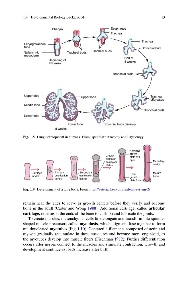
Fig. 1.8 Lung development in humans. From OpenStax: Anatomy and Physiology
Cartilage
model
Primary
ossification
centre
Growth
plates or
epiphyseal
plates
Distal
growth
plate fused
Proximal
growth
plate still
open
Medullary
cavity
Mature
bone
Secondary
ossification
centre
Fig. 1.9 Development of a long bone. From https://veteriankey.com/skeletal-system-2/
remain near the ends to serve as growth centers before they ossify and become
bone in the adult (Carter and Wong 1988). Additional cartilage, called articular
cartilage, remains at the ends of the bone to cushion and lubricate the joints.
To create muscles, mesenchymal cells first elongate and transform into spindleshaped muscle precursors called myoblasts, which align and fuse together to form
multinucleated myotubes (Fig. 1.10). Contractile filaments composed of actin and
myosin gradually accumulate in these structures and become more organized, as
the myotubes develop into muscle fibers (Fischman 1972). Further differentiation
occurs after nerves connect to the muscles and stimulate contraction. Growth and
development continue as loads increase after birth.
13
Fig. 1.8 Lung development in humans. From OpenStax: Anatomy and Physiology
Cartilage
model
Primary
ossification
centre
Growth
plates or
epiphyseal
plates
Distal
growth
plate fused
Proximal
growth
plate still
open
Medullary
cavity
Mature
bone
Secondary
ossification
centre
Fig. 1.9 Development of a long bone. From https://veteriankey.com/skeletal-system-2/
remain near the ends to serve as growth centers before they ossify and become
bone in the adult (Carter and Wong 1988). Additional cartilage, called articular
cartilage, remains at the ends of the bone to cushion and lubricate the joints.
To create muscles, mesenchymal cells first elongate and transform into spindleshaped muscle precursors called myoblasts, which align and fuse together to form
multinucleated myotubes (Fig. 1.10). Contractile filaments composed of actin and
myosin gradually accumulate in these structures and become more organized, as
the myotubes develop into muscle fibers (Fischman 1972). Further differentiation
occurs after nerves connect to the muscles and stimulate contraction. Growth and
development continue as loads increase after birth.