

238
5 Contraction
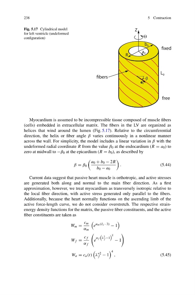
Fig. 5.17 Cylindrical model
for left ventricle (undeformed
configuration)
free
a 0
b 0
-E
Z
4
R
L 0
e F
-E
e F
Myocardium is assumed to be incompressible tissue composed of muscle fibers
(cells) embedded in extracellular matrix. The fibers in the LV are organized as
helices that wind around the lumen (Fig. 5.17). Relative to the circumferential
direction, the helix or fiber angle β varies continuously in a nonlinear manner
across the wall. For simplicity, the model includes a linear variation in β with the
undeformed radial coordinate R from the value β 0 at the endocardium (R = a 0 ) to
zero at midwall to −β 0 at the epicardium (R = b 0 ), as described by
β = β 0
a 0 + b 0 − 2R
b 0 − a 0
.
(5.44)
Current data suggest that passive heart muscle is orthotropic, and active stresses
are generated both along and normal to the main fiber direction. As a first
approximation, however, we treat myocardium as transversely isotropic relative to
the local fiber direction, with active stress generated only parallel to the fibers.
Additionally, because the heart normally functions on the ascending limb of the
active force-length curve, we do not consider overstretch. The respective strainenergy density functions for the matrix, the passive fiber constituents, and the active
fiber constituents are taken as
W m =
c m
α m
e
α m (I 1 −3)
− 1
W f =
c f
α f
e
α f
λ 2
f −1
2
− 1
W a = c a (t)
λ
∗2
f − 1
4
,
(5.45)
5 Contraction
Fig. 5.17 Cylindrical model
for left ventricle (undeformed
configuration)
free
a 0
b 0
-E
Z
4
R
L 0
e F
-E
e F
Myocardium is assumed to be incompressible tissue composed of muscle fibers
(cells) embedded in extracellular matrix. The fibers in the LV are organized as
helices that wind around the lumen (Fig. 5.17). Relative to the circumferential
direction, the helix or fiber angle β varies continuously in a nonlinear manner
across the wall. For simplicity, the model includes a linear variation in β with the
undeformed radial coordinate R from the value β 0 at the endocardium (R = a 0 ) to
zero at midwall to −β 0 at the epicardium (R = b 0 ), as described by
β = β 0
a 0 + b 0 − 2R
b 0 − a 0
.
(5.44)
Current data suggest that passive heart muscle is orthotropic, and active stresses
are generated both along and normal to the main fiber direction. As a first
approximation, however, we treat myocardium as transversely isotropic relative to
the local fiber direction, with active stress generated only parallel to the fibers.
Additionally, because the heart normally functions on the ascending limb of the
active force-length curve, we do not consider overstretch. The respective strainenergy density functions for the matrix, the passive fiber constituents, and the active
fiber constituents are taken as
W m =
c m
α m
e
α m (I 1 −3)
− 1
W f =
c f
α f
e
α f
λ 2
f −1
2
− 1
W a = c a (t)
λ
∗2
f − 1
4
,
(5.45)