

234
5 Contraction
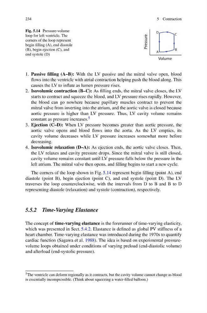
Fig. 5.14 Pressure-volume
loop for left ventricle. The
corners of the loop represent
begin filling (A), end diastole
(B), begin ejection (C), and
end systole (D)
Pressure
Volume
A
B
C
D
1. Passive filling (A–B): With the LV passive and the mitral valve open, blood
flows into the ventricle with atrial contraction helping push the blood along. This
causes the LV to inflate as lumen pressure rises.
2. Isovolumic contraction (B–C): As filling ends, the mitral valve closes, the LV
starts to contract and squeeze the blood, and LV pressure rises rapidly. However,
the blood can go nowhere because papillary muscles contract to prevent the
mitral valve from inverting into the atrium, and the aortic valve is closed because
aortic pressure is higher than LV pressure. Thus, LV cavity volume remains
constant as pressure increases. 5
3. Ejection (C–D): When LV pressure becomes greater than aortic pressure, the
aortic valve opens and blood flows into the aorta. As the LV empties, its
cavity volume decreases while LV pressure increases somewhat more before
decreasing.
4. Isovolumic relaxation (D–A): As ejection ends, the aortic valve closes. Then,
the LV relaxes and cavity pressure drops. Since the mitral valve is still closed,
cavity volume remains constant until LV pressure falls below the pressure in the
left atrium. The mitral valve then opens, and filling begins to start a new cycle.
The corners of the loop shown in Fig. 5.14 represent begin filling (point A), end
diastole (point B), begin ejection (point C), and end systole (point D). The LV
traverses the loop counterclockwise, with the intervals from D to B and B to D
representing diastole (relaxation) and systole (contraction), respectively.
5.5.2 Time-Varying Elastance
The concept of time-varying elastance is the forerunner of time-varying elasticity,
which was presented in Sect. 5.4.2. Elastance is defined as global PV stiffness of a
heart chamber. Time-varying elastance was introduced during the 1970s to quantify
cardiac function (Sagawa et al. 1988). The idea is based on experimental pressurevolume loops obtained under conditions of varying preload (end-diastolic volume)
and afterload (end-systolic pressure).
5 The ventricle can deform regionally as it contracts, but the cavity volume cannot change as blood
is essentially incompressible. (Think about squeezing a water-filled balloon.)
5 Contraction
Fig. 5.14 Pressure-volume
loop for left ventricle. The
corners of the loop represent
begin filling (A), end diastole
(B), begin ejection (C), and
end systole (D)
Pressure
Volume
A
B
C
D
1. Passive filling (A–B): With the LV passive and the mitral valve open, blood
flows into the ventricle with atrial contraction helping push the blood along. This
causes the LV to inflate as lumen pressure rises.
2. Isovolumic contraction (B–C): As filling ends, the mitral valve closes, the LV
starts to contract and squeeze the blood, and LV pressure rises rapidly. However,
the blood can go nowhere because papillary muscles contract to prevent the
mitral valve from inverting into the atrium, and the aortic valve is closed because
aortic pressure is higher than LV pressure. Thus, LV cavity volume remains
constant as pressure increases. 5
3. Ejection (C–D): When LV pressure becomes greater than aortic pressure, the
aortic valve opens and blood flows into the aorta. As the LV empties, its
cavity volume decreases while LV pressure increases somewhat more before
decreasing.
4. Isovolumic relaxation (D–A): As ejection ends, the aortic valve closes. Then,
the LV relaxes and cavity pressure drops. Since the mitral valve is still closed,
cavity volume remains constant until LV pressure falls below the pressure in the
left atrium. The mitral valve then opens, and filling begins to start a new cycle.
The corners of the loop shown in Fig. 5.14 represent begin filling (point A), end
diastole (point B), begin ejection (point C), and end systole (point D). The LV
traverses the loop counterclockwise, with the intervals from D to B and B to D
representing diastole (relaxation) and systole (contraction), respectively.
5.5.2 Time-Varying Elastance
The concept of time-varying elastance is the forerunner of time-varying elasticity,
which was presented in Sect. 5.4.2. Elastance is defined as global PV stiffness of a
heart chamber. Time-varying elastance was introduced during the 1970s to quantify
cardiac function (Sagawa et al. 1988). The idea is based on experimental pressurevolume loops obtained under conditions of varying preload (end-diastolic volume)
and afterload (end-systolic pressure).
5 The ventricle can deform regionally as it contracts, but the cavity volume cannot change as blood
is essentially incompressible. (Think about squeezing a water-filled balloon.)