

performance for water oxidation when using RuO 2 , IrO 2 , CoO x , or MoO 3
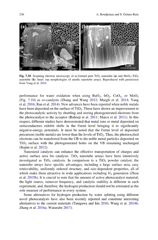
(Fig. 7.10) as co-catalysts (Zhang and Wang 2012; Maegli et al. 2014; Yang
et al. 2016; Ran et al. 2014). New advances have been reported when noble metals
have been deposited on the surface of TiO 2 . These have shown an improvement in
the photocatalytic activity by shuttling and storing photogenerated electrons from
the photocatalyst to the acceptor (Bahruji et al. 2011; Maicu et al. 2011). In this
respect, different studies have demonstrated that metal ions or metal deposited on
semiconductors exhibit shifts in the Fermi level bringing it to significantly
negative-energy potentials. It must be noted that the Fermi level of deposited
precursors (noble metals) are lower than the levels of TiO 2 . Thus, the photoexcited
electrons can be transferred from the CB to the noble metal particles deposited on
TiO 2 surface with the photogenerated holes on the VB remaining unchanged
(Rajini et al. 2012).
Nanosized catalysts can enhance the effective transportation of charges and
active surface area for catalysis. TiO 2 nanotube arrays have been intensively
investigated as TiO 2 catalysts. In comparison to a TiO 2 powder catalyst, the
nanotube arrays have specific advantages, including a large surface area, easy
retrievability, uniformly ordered structure, and size-dependent properties, all of
which make them attractive in wide applications including H 2 generation (Zhou
et al. 2015b). It is crucial to note that the amount of active photocatalyst material,
the light source, turnover frequency, and catalytic stability is different in each
experiment, and, therefore, the hydrogen production should not be estimated as the
sole measure of performance in every system.
Some alternatives for hydrogen production by water splitting using different
novel photocatalysts have also been recently reported and constitute interesting
alternatives to the current materials (Tanigawa and Irie 2016; Wang et al. 2014b;
Zhang et al. 2016a; Watanabe 2017).
Fig. 7.10 Scanning electron microscopy of as-formed pure TiO 2 nanotube (a) and MoO 3 –TiO 2
nanotube (b). Inset: top morphologies of anodic nanotube arrays. Reproduced with permission
from Yang et al. 2016
236
A. Boudjemaa and S. Gómez-Ruiz
(Fig. 7.10) as co-catalysts (Zhang and Wang 2012; Maegli et al. 2014; Yang
et al. 2016; Ran et al. 2014). New advances have been reported when noble metals
have been deposited on the surface of TiO 2 . These have shown an improvement in
the photocatalytic activity by shuttling and storing photogenerated electrons from
the photocatalyst to the acceptor (Bahruji et al. 2011; Maicu et al. 2011). In this
respect, different studies have demonstrated that metal ions or metal deposited on
semiconductors exhibit shifts in the Fermi level bringing it to significantly
negative-energy potentials. It must be noted that the Fermi level of deposited
precursors (noble metals) are lower than the levels of TiO 2 . Thus, the photoexcited
electrons can be transferred from the CB to the noble metal particles deposited on
TiO 2 surface with the photogenerated holes on the VB remaining unchanged
(Rajini et al. 2012).
Nanosized catalysts can enhance the effective transportation of charges and
active surface area for catalysis. TiO 2 nanotube arrays have been intensively
investigated as TiO 2 catalysts. In comparison to a TiO 2 powder catalyst, the
nanotube arrays have specific advantages, including a large surface area, easy
retrievability, uniformly ordered structure, and size-dependent properties, all of
which make them attractive in wide applications including H 2 generation (Zhou
et al. 2015b). It is crucial to note that the amount of active photocatalyst material,
the light source, turnover frequency, and catalytic stability is different in each
experiment, and, therefore, the hydrogen production should not be estimated as the
sole measure of performance in every system.
Some alternatives for hydrogen production by water splitting using different
novel photocatalysts have also been recently reported and constitute interesting
alternatives to the current materials (Tanigawa and Irie 2016; Wang et al. 2014b;
Zhang et al. 2016a; Watanabe 2017).
Fig. 7.10 Scanning electron microscopy of as-formed pure TiO 2 nanotube (a) and MoO 3 –TiO 2
nanotube (b). Inset: top morphologies of anodic nanotube arrays. Reproduced with permission
from Yang et al. 2016
236
A. Boudjemaa and S. Gómez-Ruiz