

4.9.1 Synthesis of Nanoporous Graphene
Synthesis of thin-layer nanoporous graphene involves two steps. The first step
involves the fabrication of two-dimensional monolayer graphene sheet followed
by the development of precise nanometer-sized pores in it. Thin-layer graphene
sheets can be synthesized by bottom–up approach like chemical reduction or top–
down approach which includes chemical vapor deposition (CVD) and electrochemical synthesis. Nanosized pores are introduced on the graphene sheet by various
techniques mentioned in previous sections where the size of the pore is too large to
allow the flow of water but too small to block the movement of ions through the
pores. Nucleation of isolated and reactive defects in graphene synthesized on copper
substrate by low-pressure CVD is performed through ion bombardment followed by
oxidative etching which allows the nucleated sites to grow into permeable pores
while retaining the structural integrity (O’Hern et al. 2014). Though CVD of
graphene is one of the preferable methods, it causes some intrinsic defects during
the deposition of graphene on copper and large cuts of size ranging from 100 nm to
200 nm during graphene transfer. In order to achieve selective blocking of defective
sites, two-scale multi-step sealing procedure is followed. Intrinsic defects are sealed
by the process called atomic layer deposition (ALD), and large defects are sealed by
interfacial polymerization reaction (O’Hern et al. 2015). In the preparation of
nanoporous graphene, creation of controlled and stable nanometer-sized pores is
technically challenging. In recent years creation of nanopores by etching has gained
immense interest among the researchers. The potential advantage of oxidative
etching technique is that the pore density and its size can be stabilized by optimizing
the etch time, which results in selective transport of ions (O’Hern et al. 2014).
Charged nanopores formed by ion etching are highly selective in allowing the
movement of ions through the pores (Sint et al. 2008). In focused electron beam
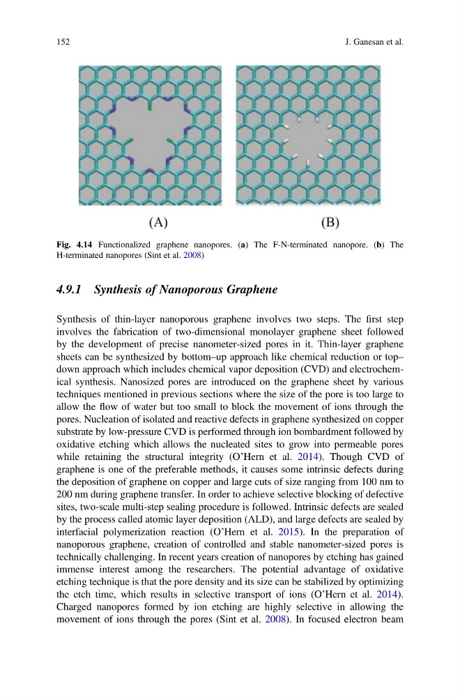
Fig. 4.14 Functionalized graphene nanopores. (a) The F-N-terminated nanopore. (b) The
H-terminated nanopores (Sint et al. 2008)
152
J. Ganesan et al.
Synthesis of thin-layer nanoporous graphene involves two steps. The first step
involves the fabrication of two-dimensional monolayer graphene sheet followed
by the development of precise nanometer-sized pores in it. Thin-layer graphene
sheets can be synthesized by bottom–up approach like chemical reduction or top–
down approach which includes chemical vapor deposition (CVD) and electrochemical synthesis. Nanosized pores are introduced on the graphene sheet by various
techniques mentioned in previous sections where the size of the pore is too large to
allow the flow of water but too small to block the movement of ions through the
pores. Nucleation of isolated and reactive defects in graphene synthesized on copper
substrate by low-pressure CVD is performed through ion bombardment followed by
oxidative etching which allows the nucleated sites to grow into permeable pores
while retaining the structural integrity (O’Hern et al. 2014). Though CVD of
graphene is one of the preferable methods, it causes some intrinsic defects during
the deposition of graphene on copper and large cuts of size ranging from 100 nm to
200 nm during graphene transfer. In order to achieve selective blocking of defective
sites, two-scale multi-step sealing procedure is followed. Intrinsic defects are sealed
by the process called atomic layer deposition (ALD), and large defects are sealed by
interfacial polymerization reaction (O’Hern et al. 2015). In the preparation of
nanoporous graphene, creation of controlled and stable nanometer-sized pores is
technically challenging. In recent years creation of nanopores by etching has gained
immense interest among the researchers. The potential advantage of oxidative
etching technique is that the pore density and its size can be stabilized by optimizing
the etch time, which results in selective transport of ions (O’Hern et al. 2014).
Charged nanopores formed by ion etching are highly selective in allowing the
movement of ions through the pores (Sint et al. 2008). In focused electron beam
Fig. 4.14 Functionalized graphene nanopores. (a) The F-N-terminated nanopore. (b) The
H-terminated nanopores (Sint et al. 2008)
152
J. Ganesan et al.